After reading this note you will be able to describe the clinical approach to SLE, recognise key tropical co‑factors relevant in Kenya, interpret the essential investigations and outline safe initial and definitive management (always verify doses and local protocols against the Kenya MoH guideline and your hospital formulary).
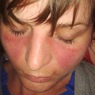
Definition
Systemic lupus erythematosus (SLE) is a chronic, multisystem autoimmune disease characterised by loss of tolerance to nuclear antigens, formation of immune complexes and complement activation, leading to episodic inflammation of skin, joints, kidneys, central nervous system, hematopoietic system and serosal membranes.
Anatomy and physiology you need
| Organ/system | Relevant structure | Why it matters in SLE |
|---|---|---|
| Skin | Epidermis & dermal basement membrane | Deposition of immune complexes → malar rash, discoid lesions |
| Joints | Synovial membrane | Non‑erosive synovitis from cytokine‑driven inflammation |
| Kidneys | Glomerular basement membrane, mesangium | Immune‑complex mediated proliferative glomerulonephritis |
| CNS | Blood‑brain barrier, cerebral vessels | Vasculitis & antibody‑mediated neuronal injury |
| Hematopoietic | Bone marrow, peripheral blood cells | Auto‑antibody mediated cytopenias |
| Serosa | Pleura, pericardium | Serositis from complement‑driven inflammation |
| Complement system | C1–C9 | Classical pathway activation amplifies tissue injury |
Causes and risk factors
- Genetic predisposition – HLA‑DR2/DR3, family clustering.
- Sex hormones – Female predominance; estrogen may enhance auto‑antibody production.
- Environmental triggers – Ultraviolet light, silica dust, smoking.
- Infections prevalent in Kenya –
- HIV – Immune dysregulation can unmask or exacerbate autoimmunity.
- Tuberculosis – Chronic antigenic stimulation; TB drugs may precipitate flares.
- Malaria – Repeated parasitaemia provides molecular mimicry for nuclear antigens.
- Chronic viral hepatitis – Hepatitis B/C can trigger immune complex formation.
- Co‑existing hematologic disorders – Sickle cell disease may alter immune regulation and mask cytopenias.
- Rheumatic fever history – Molecular mimicry between streptococcal antigens and nuclear components.
- Socio‑economic factors – Limited access to early diagnosis, delayed treatment, and higher infection burden increase morbidity.
Pathophysiology
Genetic susceptibility → loss of central/peripheral tolerance → auto‑reactive B‑cells produce anti‑nuclear antibodies (ANA, anti‑dsDNA, anti‑Sm) → formation of circulating immune complexes → deposition in tissues → activation of complement (classical pathway) → recruitment of neutrophils/macrophages → release of proteolytic enzymes and cytokines (IL‑6, TNF‑α, IFN‑α) → tissue inflammation and organ damage.
Clinical features
| Domain | Key history questions | Why they matter (pathophysiologic link) |
|---|---|---|
| General | Fatigue, fever, weight loss, night sweats | Cytokine‑driven inflammation and anemia of chronic disease produce constitutional symptoms; fever may be low‑grade and intermittent. |
| Musculoskeletal | Joint pain or swelling, morning stiffness, pattern of involvement (symmetrical, small joints), history of non‑erosive arthritis | Immune complex deposition in synovium causes transient, migratory, non‑erosive arthritis; distinguishes from rheumatoid arthritis. |
| Dermatologic | Photosensitive rash, malar (“butterfly”) rash, discoid lesions, livedo reticularis, oral or nasal ulcers, alopecia | UV‑induced apoptosis releases nuclear antigens, amplifying auto‑antibody formation; mucosal ulcers are a hallmark of vasculitis. |
| Renal | Swelling of ankles/feet, frothy urine, decreased urine output, hypertension | Lupus nephritis (immune complex glomerulonephritis) leads to proteinuria, edema, and secondary hypertension. |
| Cardiopulmonary | Chest pain (pleuritic), dyspnoea, pericardial rub, history of pleuritis or pericarditis, palpitations | Serositis from immune complex deposition in pleura/pericardium; pulmonary involvement may be hemorrhage or interstitial disease. |
| Neurologic | Headache, seizures, psychosis, mood changes, peripheral neuropathy, memory loss | Central or peripheral nervous system vasculitis; antiphospholipid antibodies can cause cerebrovascular events. |
| Hematologic | Easy bruising, bleeding, recurrent infections, history of anemia, leukopenia, thrombocytopenia | Auto‑immune cytopenias from antibody‑mediated destruction; splenic sequestration. |
| Obstetric / Reproductive | History of miscarriage, stillbirth, pre‑eclampsia, menstrual irregularities | Antiphospholipid antibodies cause placental thrombosis; hormonal dysregulation may be secondary to disease activity. |
| Infectious / Tropical context | Recent or past TB, malaria, HIV status, sickle‑cell crises, exposure to rheumatic fever | Co‑existing infections can mimic or trigger flares; immunosuppression from HIV alters presentation and management. |
Examination findings and rationale
| System | Typical finding | Pathophysiologic explanation |
|---|---|---|
| Skin | Malar rash sparing nasolabial folds, discoid plaques, photosensitivity, livedo reticularis | Cutaneous immune complex deposition; UV light increases antigen exposure. |
| Musculoskeletal | Swollen, tender small joints (MCP, PIP) without erosions, limited range of motion, “rubbery” feel | Synovitis from immune complexes; lack of erosions differentiates from erosive arthritides. |
| Cardiovascular | Pericardial friction rub, muffled heart sounds if effusion, tachycardia | Serosal inflammation; fluid accumulation can impair cardiac filling. |
| Respiratory | Pleural friction rub, decreased breath sounds at bases, crackles if alveolar hemorrhage | Pleural serositis or pulmonary capillaritis leading to hemorrhage. |
| Renal | Edema of lower limbs, hypertension, possible flank tenderness | Glomerulonephritis causing sodium retention and protein loss. |
| Neurologic | Focal deficits, cranial nerve palsies, altered mental status, seizures | Cerebral vasculitis or antiphospholipid‑mediated thrombosis. |
| Hematologic | Petechiae, ecchymoses, lymphadenopathy, splenomegaly | Cytopenias from peripheral destruction; splenic sequestration. |
| Obstetric | Uterine tenderness, signs of miscarriage (if pregnant) | Antiphospholipid antibody‑mediated placental thrombosis. |
| General | Low‑grade fever, weight loss, malaise | Systemic inflammatory response. |
Differential diagnosis
| Condition | Points toward it | Distinguishing features |
|---|---|---|
| Rheumatoid arthritis | Symmetrical small‑joint arthritis, morning stiffness | Presence of erosions on X‑ray, rheumatoid factor/anti‑CCP positivity, lack of photosensitive rash. |
| Systemic sclerosis | Skin thickening, Raynaud’s, esophageal dysmotility | Sclerodactyly, anti‑centromere or anti‑Scl‑70 antibodies, absent malar rash. |
| Dermatomyositis | Gottron papules, heliotrope rash, proximal muscle weakness | Elevated CK, muscle biopsy with perifascicular atrophy, absent renal involvement. |
| Mixed connective tissue disease | Overlap of SLE, scleroderma, polymyositis features | High anti‑U1 RNP titre, prominent Raynaud’s, less renal disease. |
| Infective endocarditis | Fever, new murmur, positive blood cultures | Vegetations on echo, embolic phenomena, no ANA/anti‑dsDNA. |
| Tuberculosis (extrapulmonary) | Fever, weight loss, pleuritis, endemic exposure | Positive GeneXpert/AFB, granulomatous histology, no ANA. |
| HIV‑associated immune complex disease | Low CD4, opportunistic infections, rash | Positive HIV serology, viral load, response to antiretrovirals. |
| Malaria (severe) | Fever spikes, hemolysis, travel history | Peripheral smear positive for parasites, rapid antigen test, no auto‑antibodies. |
| Sickle cell vaso‑occlusive crisis | Pain crises, hemolytic anemia, sickle cell trait | Hemoglobin electrophoresis positive, no serositis or rash. |
Investigations
| Test | What it answers | Interpretation |
|---|---|---|
| ANA (screen) | Presence of antinuclear antibodies | Positive in >95 % of SLE; screening test only. |
| Anti‑dsDNA, anti‑Sm | Disease‑specific auto‑antibodies | Anti‑dsDNA correlates with renal activity; anti‑Sm highly specific. |
| Complement C3, C4 | Immune complex consumption | Low levels suggest active disease, especially renal. |
| Urinalysis with microscopy | Renal involvement | Proteinuria, hematuria, cellular casts indicate lupus nephritis. |
| 24‑hour urine protein | Quantify protein loss | >0.5 g/day suggests significant nephritis; guides severity. |
| CBC with differential | Hematologic involvement | Anemia of chronic disease, leukopenia, thrombocytopenia. |
| ESR/CRP | Inflammatory activity | Elevated ESR common; CRP may be normal unless infection. |
| Renal ultrasound | Structural assessment | May show increased echogenicity; rules out obstruction. |
| Chest X‑ray | Pulmonary/pleural disease | Pleural effusion, infiltrates, cardiomegaly. |
| Echocardiogram | Pericardial effusion, valvular disease | Detects tamponade risk, Libman‑Sacks endocarditis. |
| Antiphospholipid antibody panel (LA, aCL, β2‑GP1) | Thrombotic risk | Positive in secondary antiphospholipid syndrome; guides anticoagulation. |
| Skin or renal biopsy (if indicated) | Histologic confirmation | “Full‑house” immunofluorescence (IgG, IgA, IgM, C3, C1q) in lupus nephritis. |
Diagnosis and severity
Classification (clinical + laboratory)
- Definite SLE: ≥4 of the 2019 EULAR/ACR criteria, with ANA ≥1:80 as entry criterion. Key items include constitutional, mucocutaneous, musculoskeletal, renal, neuropsychiatric, hematologic, and immunologic domains.
- Organ‑specific activity is graded separately (e.g., renal activity by proteinuria, serum creatinine, and biopsy class).
Severity grading (practical for Kenyan settings)
| Grade | Organ involvement | Clinical implication |
|---|---|---|
| Mild | Cutaneous, arthritis, serositis without organ damage | Outpatient management, NSAIDs, hydroxychloroquine. |
| Moderate | Persistent proteinuria (<1 g/day), mild cytopenias, mild neuro‑psychiatric symptoms | Add low‑dose glucocorticoids, consider immunosuppressants (azathioprine, mycophenolate). |
| Severe | Nephrotic‑range proteinuria, renal failure, CNS lupus, severe hematologic cytopenia, life‑threatening serositis | High‑dose glucocorticoids, induction with cyclophosphamide or mycophenolate, possible plasma exchange. |
| Life‑threatening | Diffuse alveolar hemorrhage, catastrophic antiphospholipid syndrome, fulminant lupus nephritis with rapid decline | Multidisciplinary ICU care, aggressive immunosuppression, anticoagulation if APS. |
Severity guides intensity of immunosuppression, need for specialist referral, and frequency of monitoring (e.g., weekly labs for severe disease vs. monthly for mild).
Management
Immediate stabilisation
- Airway‑breathing‑circulation: ensure patent airway, give supplemental O₂ if SpO₂ < 94 %, establish IV access.
- Severe organ involvement:
- Renal crisis / nephrotic syndrome – start IV methylprednisolone (pulse) and arrange urgent nephrology review.
- Neuro‑lupus (seizure, psychosis, transverse myelitis) – give IV methylprednisolone, consider plasma exchange if refractory.
- Serositis with tamponade – emergency pericardiocentesis.
- Infection screen: obtain blood, urine, sputum cultures before high‑dose steroids; give empiric broad‑spectrum antibiotics if febrile or neutropenic.
Definitive treatment (step‑wise, organ‑based)
| Goal | First‑line agents | When to add / switch | Rationale |
|---|---|---|---|
| Control systemic inflammation | Oral prednisone (taper) + hydroxychloroquine | Persistent activity after 4‑6 weeks or organ‑specific flare | Hydroxychloroquine reduces flares, improves survival; steroids give rapid control |
| Maintain remission / steroid‑sparing | Azathioprine, mycophenolate mofetil, methotrexate | Renal disease, musculoskeletal disease, steroid toxicity | Mycophenolate preferred for proliferative nephritis; azathioprine for milder disease |
| Severe refractory disease | Cyclophosphamide (IV) or rituximab | Life‑threatening renal, CNS, or vasculitic manifestations not responding to steroids + DMARD | Cyclophosphamide for class III/IV lupus nephritis; rituximab useful in refractory or when cyclophosphamide contraindicated |
| Antiphospholipid syndrome (APS) | Long‑term warfarin (target INR 2‑3) | History of thrombosis or obstetric loss | Prevents recurrent clotting; DOACs not recommended in high‑risk APS |
| Cutaneous disease | Topical steroids, calcineurin inhibitors | Persistent rash despite systemic therapy | Local control while minimizing systemic exposure |
Monitoring & safety
- Baseline: CBC, renal & liver panels, fasting glucose, lipid profile, ophthalmic exam (hydroxychloroquine).
- Follow‑up: CBC & chemistry every 4–6 weeks while on steroids/DMARDs, then every 3 months; urine protein/creatinine ratio each visit; retinal screening annually after 5 years of hydroxychloroquine.
- Adverse‑effect vigilance:
- Steroid‑induced hyperglycaemia, hypertension, osteoporosis → start calcium/vitamin D, consider bisphosphonate, monitor BP/glucose.
- Azathioprine/mycophenolate – watch for leukopenia, hepatotoxicity.
- Cyclophosphamide – monitor for haematuria, bladder toxicity; ensure hydration.
- Hydroxychloroquine – screen for retinopathy; counsel on photosensitivity.
Contra‑indications / safety notes
- Hydroxychloroquine contraindicated in known retinal disease.
- Cyclophosphamide avoided in active infection, severe cytopenia, or pregnancy (use rituximab instead).
- Warfarin avoided in uncontrolled bleeding or severe liver disease; ensure INR monitoring.
Complications and prognosis
- Renal failure (most important predictor of mortality); early aggressive immunosuppression improves long‑term renal survival.
- Neuro‑lupus – seizures, psychosis, cerebrovascular disease; may become permanent if treatment delayed.
- Cardiovascular disease – accelerated atherosclerosis; requires aggressive lipid and BP control.
- Infections – opportunistic (TB, fungal) due to immunosuppression; prophylaxis (e.g., TMP‑SMX for Pneumocystis) in high‑dose steroid regimens.
- Pregnancy complications – pre‑eclampsia, fetal loss; hydroxychloroquine is safe and reduces flare risk.
- Malignancy – modestly increased risk of non‑Hodgkin lymphoma and cervical cancer; routine screening advised.
- Mortality – historically 10‑15 % at 5 years; improved with early recognition, hydroxychloroquine use, and renal‑protective regimens.
High‑yield exam points
- Look‑alike conditions: drug‑induced lupus (hydralazine, procainamide), mixed connective tissue disease, dermatomyositis, infection‑related vasculitis.
- Commonest wrong first step: starting high‑dose steroids without ruling out active infection (especially TB, HIV, malaria in Kenya).
- Bedside examiner questions:
- “What is the significance of a positive anti‑dsDNA and low complement?”
- “How would you differentiate lupus nephritis from post‑infectious glomerulonephritis?”
- “Explain why hydroxychloroquine is continued even when the patient is in remission.”
- “What prophylaxis would you give to a patient on long‑term steroids in a malaria‑endemic area?”
Clinical case
Case: A 28‑year‑old Kenyan woman presents with a 3‑week history of joint pain (wrists, knees), photosensitive rash over cheeks, and swelling of both ankles. She reports frothy urine for 5 days. Examination shows a malar rash, oral ulcers, non‑erosive arthritis, and 2+ proteinuria on dipstick. Labs: ANA positive, anti‑dsDNA high, C3 low, creatinine 1.6 mg/dL, urine protein‑creatinine ratio 1.2 g/g.
Questions
- What is the most appropriate next step?
Answer: Initiate oral prednisone and hydroxychloroquine while arranging a renal biopsy to classify lupus nephritis.
- Which medication should be added to reduce long‑term steroid toxicity?
Answer: Start a steroid‑sparing immunosuppressant such as mycophenolate mofetil (preferred for class III/IV nephritis).
- What counseling is essential regarding future pregnancy?
Answer: Advise that disease control before conception is vital; continue hydroxychloroquine (safe in pregnancy) and avoid teratogenic drugs (e.g., cyclophosphamide).
Practice questions
Question 1
A 22‑year‑old male with known SLE presents with fever, cough, and new‑onset pleuritic chest pain. CXR shows bilateral infiltrates. What is the safest immediate action?
A. Increase prednisone dose to the protocol dose daily
B. Start empiric broad‑spectrum antibiotics and obtain sputum culture
C. Begin high‑dose IV cyclophosphamide
D. Add hydroxychloroquine
Answer: B. start empiric broad‑spectrum antibiotics and obtain sputum culture
Explanation: Infection must be ruled out before escalating immunosuppression; pleuritic pain may represent infection or serositis, but antibiotics are first‑line.
Question 2
A patient with SLE and class IV lupus nephritis is being considered for cyclophosphamide. Which finding would make you choose rituximab instead?
A. Persistent proteinuria after 6 months of therapy
B. Positive anti‑phospholipid antibodies
C. History of severe cytopenia on prior azathioprine
D. Pregnancy in the first trimester
Answer: D. pregnancy in the first trimester
Explanation: Cyclophosphamide is teratogenic; rituximab, though also not ideal, is preferred when pregnancy cannot be avoided.
Question 3
Which laboratory pattern most strongly suggests active lupus nephritis rather than a simple urinary tract infection?
A. Positive urine culture with >10⁵ CFU/mL
B. Low complement C3 and high anti‑dsDNA
C. Isolated microscopic hematuria without proteinuria
D. Elevated serum IgE
Answer: B. low complement C3 and high anti‑dsDNA
Explanation: Complement consumption and anti‑dsDNA correlate with immune complex–mediated renal inflammation.
Question 4
A 35‑year‑old woman with SLE complains of new‑onset headaches and visual disturbances. MRI is normal. Which treatment is indicated first?
A. Oral hydroxychloroquine increase
B. High‑dose IV methylprednisolone pulse
C. Start low‑dose aspirin
D. Begin antiepileptic prophylaxis
Answer: B. high‑dose IV methylprednisolone pulse
Explanation: Neuro‑lupus requires rapid high‑dose steroids to prevent irreversible damage.
Question 5
Which of the following is NOT a recommended prophylactic measure for a patient on long‑term steroids living in a malaria‑endemic region?
A. Insecticide‑treated bed nets
B. Intermittent preventive treatment with sulfadoxine‑pyrimethamine
C. Daily chloroquine prophylaxis
D. Seasonal malaria chemoprevention with amodiaquine‑pyrimethamine
Answer: C. daily chloroquine prophylaxis
Explanation: Chloroquine resistance is widespread in Kenya; it is not recommended for prophylaxis.
Drafted with AI assistance from standard references and reviewed for structure. Check details against your lecture notes and the Kenya MoH guideline.