Year 4 MBChB — Internal Medicine | Neurology foundation notes
After studying this note, you should be able to describe the main CNS structures and pathways, recognise the clinical effects of lesions at different levels, perform a structured neurological assessment, and select appropriate initial investigations.
1. Definition and organisation
The central nervous system (CNS) consists of the brain and spinal cord. It receives sensory information, integrates it and coordinates motor, autonomic and higher mental functions.
- CNS: brain and spinal cord.
- Peripheral nervous system (PNS): cranial nerves, spinal nerves, nerve roots, plexuses and peripheral nerves outside the CNS.
- Autonomic nervous system: regulates involuntary functions. Its sympathetic, parasympathetic and enteric components interact with the CNS and PNS.
2. Main structures of the brain

Diagram: Cerebral lobes, brainstem and cerebellum. Source: Wikimedia Commons, Brainlobes.svg (public domain).
Cerebrum
The cerebrum has two hemispheres connected by the corpus callosum. The cerebral cortex supports voluntary movement, sensation, language, memory, planning and behaviour.
| Region | Main function | Typical lesion findings |
|---|---|---|
| Frontal lobe | Voluntary movement, planning, behaviour; speech production in the dominant hemisphere | Contralateral weakness, personality change, expressive aphasia |
| Parietal lobe | Sensory integration and spatial awareness | Contralateral sensory loss, neglect, impaired cortical sensation |
| Temporal lobe | Hearing, memory and language comprehension | Memory impairment, receptive aphasia |
| Occipital lobe | Vision | Visual field defects |
| Insula | Taste, visceral sensation and integration of internal bodily signals | Complex sensory or autonomic disturbances |
The primary motor cortex lies in the precentral gyrus of the frontal lobe. The primary somatosensory cortex lies in the postcentral gyrus of the parietal lobe.
Cerebellum
Functions:
- Coordinates the accuracy, timing and smoothness of movement.
- Maintains balance and posture.
- Contributes to motor learning and regulation of muscle tone.
Lesions may cause ipsilateral limb ataxia, dysmetria, intention tremor, nystagmus or a broad-based gait. Coordination may be impaired even when muscle power is preserved.
Brainstem
The brainstem consists of the midbrain, pons and medulla oblongata.
- Midbrain: eye movements, pupillary reflexes and motor pathways.
- Pons: connections with the cerebellum, facial functions and respiratory regulation.
- Medulla: vital cardiovascular and respiratory centres, swallowing and several cranial nerve nuclei.
Brainstem lesions may cause cranial nerve abnormalities with limb weakness, altered consciousness or life-threatening respiratory and cardiovascular dysfunction.
Thalamus, hypothalamus and basal ganglia
- Thalamus: major relay for most sensory information reaching the cerebral cortex; also participates in motor and consciousness-related networks.
- Hypothalamus: regulates temperature, hunger, thirst, circadian rhythms and autonomic functions; controls endocrine activity largely through the pituitary gland.
- Basal ganglia: regulate movement selection and control. Parkinson disease is associated with loss of dopaminergic neurons in the substantia nigra and altered basal ganglia circuits; Huntington disease commonly causes chorea and progressive cognitive and psychiatric changes.
3. Meninges and cerebrospinal fluid (CSF)

Diagram: Dura mater, arachnoid mater and pia mater. Source: Wikimedia Commons, Meninges-en.svg (CC BY-SA 3.0).
The brain and spinal cord are covered by three meninges:
- Dura mater: tough outer layer.
- Arachnoid mater: middle membrane.
- Pia mater: delicate inner membrane closely adherent to the CNS surface.
The subarachnoid space, between arachnoid and pia, contains CSF and blood vessels.
Clinical correlations:
- Epidural haematoma: blood between skull and dura; often associated with arterial injury.
- Subdural haematoma: blood between dura and arachnoid; commonly due to torn bridging veins.
- Subarachnoid haemorrhage: bleeding into the subarachnoid space; classically presents with a sudden, severe headache.
- Meningitis: inflammation of the meninges.
CSF production and circulation
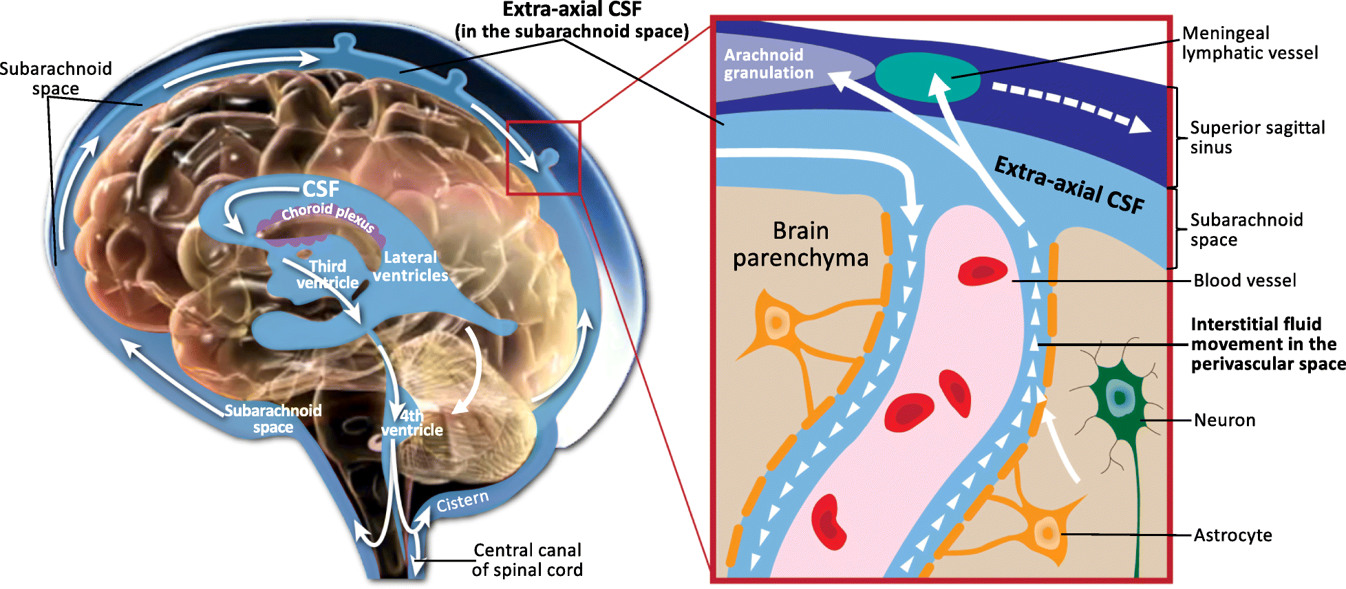
Diagram: CSF circulation and outflow. Source: Wikimedia Commons, CSF circulation.png (CC BY 4.0).
CSF is produced mainly by the choroid plexuses in the ventricles.
Flow: lateral ventricles → interventricular foramina (of Monro) → third ventricle → cerebral aqueduct → fourth ventricle → apertures of Magendie and Luschka → subarachnoid space → absorption mainly through arachnoid granulations into venous blood.
Functions include buoyancy and mechanical protection, maintenance of the chemical environment and clearance of substances. Adult CSF volume is approximately 150 mL, with production around 500 mL/day.
Obstruction of CSF flow or impaired absorption can cause hydrocephalus and, in some cases, raised intracranial pressure.
4. Neurons and nerve impulses
A neuron is a specialised signalling cell.
- Cell body: contains the nucleus and supports metabolism.
- Dendrites: receive signals.
- Axon: conducts impulses away from the cell body.
- Myelin sheath: increases conduction speed.
- Synapse: junction through which a neuron communicates with another cell.
Action potential
- At rest, ion gradients and membrane permeability maintain the resting membrane potential.
- Depolarisation usually occurs when voltage-gated sodium channels open and sodium enters.
- Repolarisation mainly occurs as potassium leaves through voltage-gated potassium channels.
- The membrane returns towards its resting state.
In myelinated axons, saltatory conduction allows impulses to be regenerated at the nodes of Ranvier, increasing conduction speed.
Important neurotransmitters
| Neurotransmitter | Main associations |
|---|---|
| Acetylcholine | Neuromuscular transmission, autonomic function and cognition |
| Dopamine | Movement, motivation and reward |
| Serotonin | Mood, sleep and other functions |
| GABA | Major inhibitory neurotransmitter in the brain |
| Glutamate | Major excitatory neurotransmitter in the brain |
| Noradrenaline | Alertness and sympathetic activity |
Clinical links: altered dopamine signalling contributes to Parkinsonian motor symptoms; reduced inhibitory control can contribute to seizure generation. Acetylcholinesterase inhibitors are used in myasthenia gravis and, in a different context, Alzheimer disease.
5. Motor pathways and upper versus lower motor neuron lesions
Corticospinal tract
The corticospinal tract carries commands for voluntary movement from the cerebral cortex to spinal motor circuits.
Simplified pathway: motor cortex → corona radiata → internal capsule → cerebral peduncle → pons → medullary pyramids.
Most fibres cross at the pyramidal decussation in the caudal medulla, then descend in the lateral corticospinal tract.
- A lesion above the crossing usually causes weakness on the opposite side of the body.
- A lesion below the crossing in the spinal cord usually causes weakness on the same side below the lesion.
UMN versus LMN
| Feature | Upper motor neuron (UMN) lesion | Lower motor neuron (LMN) lesion |
|---|---|---|
| Site | Motor cortex or descending motor pathways | Anterior horn cell, motor root or peripheral motor nerve |
| Weakness | Present | Present |
| Tone | Usually increased after the acute phase | Reduced |
| Deep tendon reflexes | Usually increased | Reduced or absent |
| Plantar response | May be extensor (Babinski sign) | Usually flexor or absent |
| Muscle wasting | Usually mild from disuse | Often prominent |
| Fasciculations | Not typical | May occur |
Important: an acute severe UMN lesion, such as an acute stroke, can initially cause reduced tone and reflexes before spasticity develops.
Clinical localisation examples
- Right facial and right arm/leg weakness with aphasia in a right-handed patient suggests a left hemispheric lesion.
- Weakness of both legs with a sensory level suggests a spinal cord lesion.
- Distal weakness with wasting, fasciculations and reduced reflexes suggests an LMN disorder.
- Ataxia with relatively preserved power suggests cerebellar dysfunction, although other causes must be considered.
6. Sensory pathways
Dorsal column–medial lemniscus pathway
Carries vibration, joint-position sense and discriminative (fine) touch. Fibres ascend on the same side in the spinal cord and cross in the medulla.
Spinothalamic tract
Carries pain, temperature and much of crude touch. Fibres usually cross within the spinal cord, near the level at which they enter.
Clinical application: a spinal cord lesion may affect the pathways differently. In a hemisection, vibration and position sense may be lost on the same side below the lesion, while pain and temperature may be lost on the opposite side beginning slightly below it.
7. Cranial nerves: clinical summary
| Nerve | Main function |
|---|---|
| I — Olfactory | Smell |
| II — Optic | Vision |
| III — Oculomotor | Most eye movements, eyelid elevation and pupillary constriction |
| IV — Trochlear | Superior oblique muscle |
| V — Trigeminal | Facial sensation and muscles of mastication |
| VI — Abducens | Lateral rectus; abducts the eye |
| VII — Facial | Facial expression; taste from anterior two-thirds of tongue |
| VIII — Vestibulocochlear | Hearing and balance |
| IX — Glossopharyngeal | Swallowing, posterior-third tongue taste and gag reflex afferent limb |
| X — Vagus | Palatal and laryngeal function; parasympathetic functions |
| XI — Accessory | Sternocleidomastoid and trapezius |
| XII — Hypoglossal | Tongue movement |
Clinical pearls
- CN III palsy may cause ptosis and a dilated pupil, depending on the cause.
- CN VI palsy impairs abduction of the affected eye.
- An LMN facial nerve lesion affects the ipsilateral upper and lower face.
- A typical hemispheric UMN facial lesion predominantly affects the contralateral lower face, with relative forehead sparing.
- A hypoglossal LMN lesion causes the tongue to deviate towards the affected side on protrusion.
8. Autonomic nervous system
- Sympathetic division (“fight or flight”): increases heart rate, promotes pupil dilation, generally reduces gastrointestinal motility and supports sweating and other stress responses.
- Parasympathetic division (“rest and digest”): generally reduces heart rate, promotes gastrointestinal motility and secretion, and contributes to pupil constriction and accommodation.
Autonomic dysfunction may cause orthostatic hypotension, abnormal sweating, bowel or bladder dysfunction and other involuntary-function abnormalities.
9. Neurological history and examination
Focused history
Establish:
- Onset: sudden or gradual?
- Duration, progression and previous episodes.
- Weakness, numbness, speech or visual changes.
- Headache, seizures, fever or altered consciousness.
- Trauma, medications, alcohol and vascular risk factors.
- Relevant systemic disease, infection exposure and functional impact.
Examination
- General assessment and vital signs.
- Level of consciousness and cognition.
- Cranial nerves.
- Motor system: inspection, tone, power, reflexes and plantar response.
- Sensory system.
- Coordination and gait, if safe.
- Meningeal signs when indicated.
Glasgow Coma Scale (GCS)
| Component | Score |
|---|---|
| Eye opening (E) | 1–4 |
| Verbal response (V) | 1–5 |
| Motor response (M) | 1–6 |
| Total | 3–15 |
Record the individual components, not only the total. A GCS of 8 or less indicates severe impairment of consciousness and requires urgent assessment, including airway protection. The score alone does not determine every airway decision.
10. Investigations in neurological disease
Select investigations according to the suspected diagnosis:
- Bedside glucose: essential in altered consciousness or suspected stroke.
- Full blood count: evaluates anaemia, infection and other haematological abnormalities.
- Electrolytes, renal function and calcium: assess metabolic causes of neurological symptoms or seizures.
- CT brain: useful for suspected acute haemorrhage, trauma and selected acute neurological presentations.
- MRI brain or spine: detailed assessment of many parenchymal, demyelinating and spinal cord disorders.
- Lumbar puncture: selected suspected meningitis, encephalitis or other CSF disorders. Assess contraindications first; imaging is needed before LP in selected patients at risk of dangerous intracranial pressure gradients.
- EEG: evaluates suspected seizures and selected disorders of consciousness.
- Nerve conduction studies and EMG: assess peripheral nerve and motor-unit disorders.
11. Emergency principles
For acute neurological deterioration:
- Assess airway, breathing and circulation.
- Check oxygenation, vital signs and bedside glucose.
- In suspected stroke, establish the exact symptom-onset time or last-known-well time.
- Identify immediately reversible causes, including hypoglycaemia.
- Arrange urgent appropriate imaging and specialist assessment if stroke, haemorrhage or another neurological emergency is suspected.
- Treat seizures, infection, raised intracranial pressure or other emergencies according to the presentation and local protocols.
Emergency warning: sudden facial weakness, arm weakness or speech disturbance should be treated as possible stroke and requires immediate emergency assessment.
12. CAT practice questions
1. A patient has right-sided weakness and aphasia. Where is the likely lesion in a right-handed patient?
Answer: The left cerebral hemisphere, commonly involving the dominant hemisphere's motor pathways and language areas.
2. A patient has spasticity, hyperreflexia and an extensor plantar response. Is this an UMN or LMN lesion?
Answer: UMN lesion.
3. Which pathway carries vibration and joint-position sense?
Answer: Dorsal column–medial lemniscus pathway.
4. A patient has ipsilateral limb ataxia but preserved power. Which structure is most likely affected?
Answer: The cerebellum or its connections.
5. What bedside test must be checked promptly in a patient with altered consciousness?
Answer: Blood glucose, alongside immediate assessment of airway, breathing and circulation.
6. What are the three components of the GCS?
Answer: Eye opening, verbal response and motor response.
7. Why should the exact onset time be established in suspected stroke?
Answer: It affects eligibility for time-dependent reperfusion treatment and other urgent decisions.
Key takeaways
- Localise the lesion before deciding the cause.
- UMN lesions typically cause increased tone and reflexes after the acute phase; LMN lesions typically cause reduced tone and reflexes.
- Motor fibres mostly cross in the caudal medulla; pain and temperature fibres cross near their spinal entry level.
- Sudden focal neurological deficits are emergencies.
- Choose investigations based on the clinical presentation, and always check glucose early in altered consciousness.