After this note, you should be able to define pulmonary hypertension and cor pulmonale, describe their causes in the Kenyan context, and outline their pathophysiology. Always check drug doses and local protocols against the Kenya MoH guidelines and the hospital formulary.
Definition
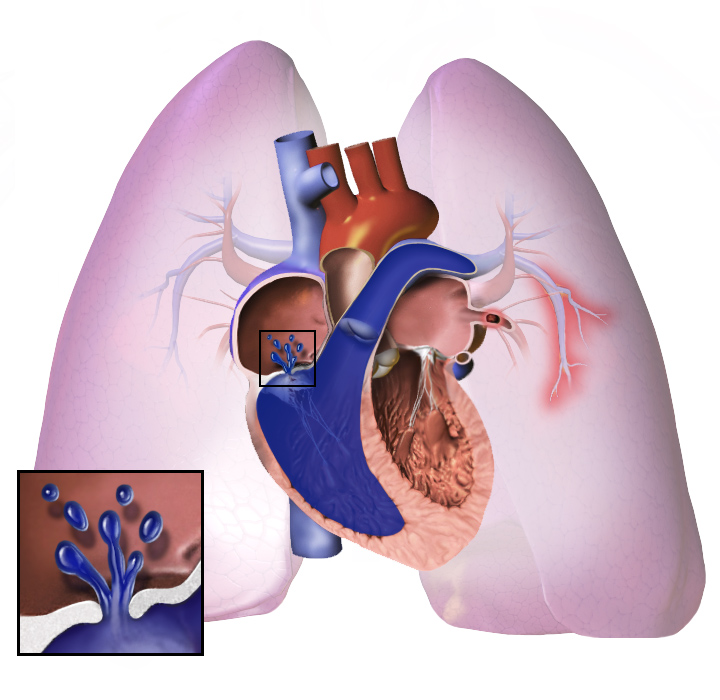
Pulmonary Hypertension (PH) is a haemodynamic and pathophysiological condition defined by an increase in mean pulmonary arterial pressure (mPAP) ≥ 20 mmHg at rest, as measured by right heart catheterisation (RHC). This elevated pressure is due to a combination of increased pulmonary vascular resistance (PVR), increased pulmonary blood flow, and/or increased left atrial pressure.
Cor Pulmonale is right ventricular (RV) enlargement (hypertrophy, dilatation, or both) due to PH caused by primary disorders of the lung parenchyma, pulmonary vasculature, or disorders of breathing. It is a diagnosis of exclusion, meaning PH due to left heart disease or congenital heart disease must be ruled out.
Anatomy and physiology you need
The pulmonary circulation is a low-pressure, high-flow system.
- Pulmonary arteries: Carry deoxygenated blood from the right ventricle to the lungs. They are thin-walled and highly compliant compared to systemic arteries.
- Pulmonary capillaries: Site of gas exchange.
- Pulmonary veins: Carry oxygenated blood from the lungs to the left atrium.
- Right ventricle (RV): A thin-walled chamber adapted to pump against low resistance. Chronic increases in pulmonary vascular resistance lead to RV hypertrophy and eventual dilatation and failure.
- Pulmonary vascular resistance (PVR): Determined by the radius of the pulmonary arterioles. Vasoconstriction, remodelling, or obstruction increases PVR.
- Pulmonary blood flow: Normally equal to cardiac output.
- Pulmonary arterial pressure (PAP): A function of pulmonary blood flow, PVR, and left atrial pressure.
Causes and risk factors
The World Health Organization (WHO) classifies PH into five groups based on shared pathophysiological mechanisms, clinical presentation, and therapeutic approaches.
| WHO Group | Category | Common Causes in Kenya |
|---|---|---|
| Group 1 | Pulmonary Arterial Hypertension (PAH) | Idiopathic, HIV, portal hypertension, schistosomiasis, congenital heart disease (e.g., VSD, PDA, ASD with Eisenmenger's) |
| Group 2 | PH due to Left Heart Disease | Left ventricular systolic or diastolic dysfunction, valvular heart disease (mitral stenosis, aortic stenosis/regurgitation) |
| Group 3 | PH due to Lung Diseases and/or Hypoxia | COPD, interstitial lung disease (ILD), chronic high altitude exposure, obstructive sleep apnoea, TB sequelae (fibrosis, bronchiectasis) |
| Group 4 | Chronic Thromboembolic PH (CTEPH) | Previous pulmonary embolism (PE), recurrent DVT, antiphospholipid syndrome |
| Group 5 | PH with Unclear Multifactorial Mechanisms | Haematological disorders (sickle cell disease, myeloproliferative disorders), systemic disorders (sarcoidosis, chronic renal failure), metabolic disorders (glycogen storage diseases), external compression of pulmonary vasculature |
Specific causes in our Kenyan setting:
- Chronic Lung Disease: COPD (due to biomass fuel exposure, smoking), interstitial lung disease (e.g., post-TB fibrosis, pneumoconiosis), severe asthma.
- Schistosomiasis: Schistosoma mansoni eggs embolise to the pulmonary arteries, causing granuloma formation, inflammation, and vascular remodelling.
- Thromboembolism: Recurrent or unresolved pulmonary emboli leading to CTEPH. Risk factors include DVT, prolonged immobility, hypercoagulable states.
- Left Heart Disease: Rheumatic heart disease (especially mitral stenosis), hypertensive heart disease leading to left ventricular dysfunction.
- Sickle Cell Disease: Chronic haemolysis, endothelial dysfunction, and vaso-occlusion contribute to PH.
- HIV Infection: Direct viral effects on pulmonary vasculature, increased inflammation, and opportunistic infections.
- Congenital Heart Disease: Uncorrected shunts (e.g., VSD, PDA) leading to increased pulmonary blood flow and eventual Eisenmenger's syndrome.
Pathophysiology
The underlying pathophysiology varies by WHO group, but the common endpoint is increased pulmonary arterial pressure and subsequent right ventricular strain.
General Pathophysiology: Increased PVR OR Increased Pulmonary Blood Flow OR Increased Left Atrial Pressure ↓ Increased Pulmonary Arterial Pressure ↓ Increased Right Ventricular Afterload ↓ Right Ventricular Hypertrophy (compensatory) ↓ Right Ventricular Dilatation and Dysfunction (decompensatory) ↓ Right Heart Failure (Cor Pulmonale)
Specific Mechanisms:
- Hypoxia (e.g., Group 3 PH):
Alveolar Hypoxia → Pulmonary Arteriolar Vasoconstriction (protective in acute settings, maladaptive chronically) → Increased PVR → PH
- Vascular Remodelling (e.g., Group 1 PAH, Schistosomiasis):
Endothelial Dysfunction + Smooth Muscle Proliferation + Fibrosis + In situ Thrombosis → Narrowing and Obstruction of Pulmonary Arterioles → Increased PVR → PH
- Thromboembolism (Group 4 CTEPH):
Unresolved Pulmonary Emboli → Organisation and Fibrosis within Pulmonary Arteries → Fixed Obstruction and Remodelling of Unobstructed Vessels → Increased PVR → PH
- Left Heart Disease (Group 2 PH):
Increased Left Atrial Pressure (e.g., due to mitral stenosis or LV failure) → Backward Transmission of Pressure to Pulmonary Veins and Capillaries → Pulmonary Venous Hypertension → Reactive Pulmonary Arteriolar Vasoconstriction and Remodelling → PH
Clinical features
History questions that matter:
- Dyspnoea: Onset, progression (exertional, then at rest), severity (NYHA class), impact on daily activities. This is the cardinal symptom, reflecting reduced cardiac output and impaired gas exchange.
- Fatigue/Weakness: Due to reduced cardiac output and tissue hypoperfusion.
- Chest pain: Anginal type (right ventricular ischaemia due to increased wall tension) or pleuritic (pulmonary infarction, RV distension).
- Syncope/Pre-syncope: Exertional syncope is a red flag, indicating severe PH and inability to increase cardiac output adequately, leading to cerebral hypoperfusion.
- Palpitations: Arrhythmias (atrial fibrillation, ventricular ectopy) due to RV strain and dilation.
- Peripheral oedema/Abdominal distension: Symptoms of right heart failure (fluid retention, ascites).
- Haemoptysis: Due to rupture of dilated pulmonary vessels or pulmonary infarction.
- Raynaud's phenomenon: Associated with connective tissue diseases (e.g., scleroderma) that can cause PH.
- Family history: Of PH or sudden death (heritable PH).
- Exposure history: Schistosomiasis, HIV, illicit drug use (anorexigens).
- Symptoms of underlying conditions: Cough, wheeze (COPD, asthma), orthopnoea, PND (left heart disease), DVT/PE history (CTEPH).
Examination findings and why each occurs:
- General appearance: Cyanosis (hypoxia), cachexia (severe chronic illness), jaundice (hepatic congestion).
- Vital signs: Tachycardia (compensatory, arrhythmias), tachypnoea (dyspnoea), hypotension (severe RV failure).
- Jugular Venous Pressure (JVP): Elevated, often with a prominent 'a' wave (right atrial contraction against a stiff RV) and sometimes a prominent 'v' wave (tricuspid regurgitation). Reflects increased right atrial pressure.
- Precordial palpation:
- Left parasternal heave: Right ventricular hypertrophy and dilation.
- Palpable P2: Loud pulmonary component of S2, indicating increased pulmonary artery pressure.
- Auscultation:
- Loud P2: Pathognomonic for PH, due to forceful closure of the pulmonary valve.
- Right-sided S3: Right ventricular dysfunction/failure.
- Right-sided S4: Right ventricular hypertrophy/stiffness.
- Pansystolic murmur at lower left sternal border: Tricuspid regurgitation (due to RV dilation).
- Early diastolic murmur at upper left sternal border: Pulmonary regurgitation (Graham Steell murmur, due to pulmonary artery dilation).
- Crackles/wheezes: Underlying lung disease.
- Abdomen: Hepatomegaly (tender, pulsatile), ascites – signs of right heart failure and hepatic congestion.
- Extremities: Peripheral oedema (pitting, bilateral) – right heart failure. Clubbing (chronic hypoxia, especially with underlying lung disease or congenital heart disease with shunt).
Differential diagnosis
| Condition | What points to it | What separates it from PH |
|---|---|---|
| Bronchial Asthma | Episodic wheeze, cough, reversibility | Normal P2, no RV strain signs, spirometry confirms obstruction |
| Congestive Heart Failure | Orthopnoea, PND, S3 gallop, bilateral crackles | Predominant left heart failure signs, echo shows LV dysfunction |
| Chronic Obstructive Lung Disease | Chronic cough, sputum, smoking history, barrel chest | Spirometry shows irreversible obstruction, often mild PH |
| Interstitial Lung Disease | Progressive dyspnoea, dry cough, fine crackles | HRCT shows characteristic fibrosis, often mild PH |
| Anaemia | Pallor, fatigue, low Hb | No RV strain signs, PH symptoms resolve with correction of anaemia |
| Anxiety/Panic Disorder | Hyperventilation, chest tightness, no physical signs | No objective evidence of PH or cardiac/pulmonary pathology |
Investigations
| Test | What it answers | What a result means |
|---|---|---|
| ECG | Right heart strain, hypertrophy, arrhythmias | RVH (tall R in V1, deep S in V6), right axis deviation, RAE |
| Chest X-ray | Cardiac chamber enlargement, pulmonary artery size, lung parenchyma | Enlarged pulmonary arteries, RV enlargement, clear lung fields (PAH) or signs of lung disease |
| Echocardiography | RV size/function, estimated PAP, valve function, shunts | RV dilation/hypertrophy, TR jet velocity (est. sPAP), D-shaped LV, pericardial effusion |
| Lung Function Tests | Underlying obstructive/restrictive lung disease | Obstructive (FEV1/FVC < 0.7), restrictive (low TLC), reduced DLCO |
| CT Pulmonary Angiogram | Pulmonary embolism, chronic thromboembolic disease, lung parenchyma | Filling defects (PE), web/bands (CTEPH), interstitial lung disease |
| V/Q Scan | Chronic thromboembolic pulmonary hypertension (CTEPH) | Mismatched perfusion defects (high probability for CTEPH) |
| Right Heart Catheterisation | Confirms PH, measures pressures, CO, PVR, vasoreactivity | mPAP ≥ 20 mmHg, PCWP ≤ 15 mmHg (pre-capillary PH), vasoreactivity testing |
Diagnosis and severity
The definitive diagnosis of pulmonary hypertension requires right heart catheterisation (RHC).
Diagnostic criteria for PH (WHO definition):
- Mean pulmonary arterial pressure (mPAP) ≥ 20 mmHg at rest, measured by RHC.
Classification based on RHC:
- Pre-capillary PH:
- mPAP ≥ 20 mmHg
- Pulmonary artery wedge pressure (PAWP) ≤ 15 mmHg
- Pulmonary vascular resistance (PVR) > 2 Wood units
- This includes Group 1 (PAH), Group 3 (PH due to lung disease), Group 4 (CTEPH), and some Group 5.
- Post-capillary PH:
- mPAP ≥ 20 mmHg
- PAWP > 15 mmHg
- This is typically Group 2 (PH due to left heart disease).
Severity assessment:
Severity is assessed by a combination of clinical symptoms, functional capacity, haemodynamics, and markers of RV function. This guides treatment decisions and prognosis.
- WHO Functional Class (FC):
- FC I: No limitation of physical activity. Ordinary physical activity does not cause undue dyspnoea or fatigue, chest pain or near syncope.
- FC II: Slight limitation of physical activity. Comfortable at rest. Ordinary physical activity results in undue dyspnoea or fatigue, chest pain or near syncope.
- FC III: Marked limitation of physical activity. Comfortable at rest. Less than ordinary activity causes undue dyspnoea or fatigue, chest pain or near syncope.
- FC IV: Inability to carry out any physical activity without symptoms. Manifests signs of right heart failure. Dyspnoea and/or fatigue may be present at rest. Discomfort is increased by any physical activity.
- 6-minute walk distance (6MWD): A measure of functional capacity. Shorter distances indicate more severe disease.
- Haemodynamics from RHC: Higher mPAP, higher PVR, lower cardiac index, higher right atrial pressure indicate more severe disease.
- Echocardiographic findings: Degree of RV dilation and dysfunction, pericardial effusion.
- Biomarkers: Elevated NT-proBNP/BNP (reflects RV stretch and dysfunction).
Management
Management of pulmonary hypertension (PH) and cor pulmonale focuses on treating the underlying cause, reducing pulmonary artery pressure, improving right ventricular function, and alleviating symptoms.
Immediate Stabilisation:
- Oxygen therapy: Crucial for hypoxemic patients to reduce pulmonary vasoconstriction and improve oxygen delivery.
- Diuretics: Cautious use to manage fluid overload and peripheral oedema, but avoid aggressive diuresis which can reduce preload and worsen right ventricular output.
- Inotropes: May be considered in severe right ventricular failure with cardiogenic shock (e.g., dobutamine) to improve cardiac output.
- Vasodilators: Systemic vasodilators (e.g., nitrates) are generally contraindicated as they can cause systemic hypotension and worsen right ventricular function by reducing preload.
Definitive Treatment:
- Treating the Underlying Cause:
- Chronic Lung Disease (e.g., COPD, ILD): Optimise bronchodilators, steroids, antibiotics for exacerbations. Long-term oxygen therapy is key for chronic hypoxemia.
- Schistosomiasis: Praziquantel to treat the parasitic infection.
- Thromboembolism (Chronic Thromboembolic PH - CTEPH): Anticoagulation is standard. Surgical pulmonary endarterectomy is curative in eligible patients. Balloon pulmonary angioplasty is an option for non-surgical candidates.
- Left Heart Disease: Optimise management of heart failure with preserved or reduced ejection fraction, valvular heart disease.
- Sickle Cell Disease: Hydroxyurea, regular transfusions, and management of vaso-occlusive crises.
- Pulmonary Vasodilator Therapy (for Group 1 PAH):
- Endothelin Receptor Antagonists (ERAs): e.g., bosentan, ambrisentan. Block endothelin-1, a potent vasoconstrictor.
- Phosphodiesterase-5 (PDE5) Inhibitors: e.g., sildenafil, tadalafil. Increase cGMP, leading to vasodilation.
- Prostacyclin Analogues: e.g., epoprostenol (IV), treprostinil (IV, SC, inhaled), iloprost (inhaled). Potent vasodilators and anti-proliferative agents.
- Guanylate Cyclase Stimulators: e.g., riociguat. Directly stimulate soluble guanylate cyclase, increasing cGMP.
- Anticoagulation: Considered for Group 1 PAH, especially if right heart catheterisation shows low cardiac output or in CTEPH.
- Diuretics: Loop diuretics (e.g., furosemide) for fluid overload, with careful monitoring of renal function and electrolytes.
- Digoxin: May be considered in severe right ventricular failure with atrial fibrillation to control heart rate and improve contractility.
Monitoring and Follow-up: Regular clinical assessment, echocardiography, 6-minute walk test, and review of symptoms.
Contraindications and Safety:
- Systemic Vasodilators: Generally avoided due to risk of systemic hypotension.
- Aggressive Diuresis: Can compromise right ventricular preload and cardiac output.
- Non-selective Beta-blockers: Can worsen right ventricular function and are generally avoided in advanced PH.
Complications and Prognosis
Complications:
- Right Heart Failure: Progressive worsening of right ventricular function, leading to severe oedema, ascites, and anasarca.
- Arrhythmias: Atrial fibrillation, atrial flutter, and ventricular arrhythmias due to right atrial and ventricular stretch and remodelling.
- Thromboembolism: Increased risk of pulmonary embolism due to stasis in the dilated right heart chambers.
- Syncope: Due to transient reduction in cardiac output, especially during exertion.
- Sudden Cardiac Death: Often due to arrhythmias or acute right heart failure.
- Renal Impairment: Due to reduced cardiac output and systemic congestion.
- Liver Congestion: Leading to impaired liver function and jaundice.
Prognosis: Prognosis in PH and cor pulmonale is variable and depends heavily on the underlying cause, the severity of PH, and the response to treatment. Untreated severe PH has a poor prognosis. Early diagnosis and targeted therapy, especially for Group 1 PAH and CTEPH, can significantly improve outcomes and quality of life. Patients with PH secondary to left heart disease or lung disease generally have a prognosis dictated by the primary condition.
High-yield exam points
- Loud P2: Signifies increased pulmonary artery pressure causing forceful closure of the pulmonary valve.
- Parasternal heave: Palpable impulse over the left sternal border, indicating right ventricular hypertrophy.
- Raised JVP: Reflects increased right atrial pressure due to right heart failure.
- Diuretics used carefully: Aggressive diuresis can reduce right ventricular preload, worsening cardiac output in a pressure-overloaded right ventricle. Aim for euvolemia, not hypovolemia.
- ECG findings: Right axis deviation, RVH (tall R in V1, deep S in V6), right atrial enlargement (P-pulmonale).
- Chest X-ray: Prominent pulmonary arteries, right ventricular enlargement (boot-shaped heart in severe cases), clear lung fields (unless underlying lung disease).
- Echocardiography: Estimation of PASP, RV size and function, tricuspid regurgitation.
- Right heart catheterisation: Gold standard for diagnosis and severity assessment, measuring PAP, PCWP, CO.
- Long-term oxygen therapy: Crucial for hypoxemic patients to reduce pulmonary vasoconstriction and improve survival in chronic lung disease.
Clinical case
A 55-year-old male presents with progressive shortness of breath, fatigue, and lower limb swelling for 6 months. He has a 30-pack-year smoking history and chronic cough. On examination, he has a raised JVP, a loud P2, a parasternal heave, and bilateral pitting oedema to the knees. His oxygen saturation is 88% on room air.
- What is the most likely underlying cause of his pulmonary hypertension?
- Model Answer: Chronic Obstructive Pulmonary Disease (COPD) leading to chronic hypoxemia and cor pulmonale. His smoking history and chronic cough are key indicators.
- What ECG findings would you expect in this patient?
- Model Answer: Right axis deviation, signs of right ventricular hypertrophy (e.g., tall R wave in V1, deep S wave in V6), and possibly P-pulmonale (peaked P waves in leads II, III, aVF) indicating right atrial enlargement.
- Why should diuretics be used cautiously in this patient?
- Model Answer: Aggressive diuresis can reduce right ventricular preload. In a patient with a pressure-overloaded right ventricle, a reduction in preload can lead to a significant drop in cardiac output and systemic hypotension, worsening his clinical condition.
Practice questions
Question 1
A 45-year-old woman with known systemic sclerosis presents with progressive dyspnea and fatigue. Echocardiography reveals severe pulmonary hypertension. Which of the following is the most appropriate initial management step for her pulmonary hypertension?
A. Aggressive diuresis with high-dose furosemide B. Initiation of a pulmonary vasodilator (e.g., sildenafil) C. Long-term systemic anticoagulation with warfarin D. Surgical pulmonary endarterectomy
Answer: B. Initiation of a pulmonary vasodilator (e.g., sildenafil) Explanation: Systemic sclerosis is a common cause of Group 1 Pulmonary Arterial Hypertension (PAH), for which pulmonary vasodilators are the mainstay of treatment.
Question 2
A 60-year-old man with severe COPD and chronic hypoxemia develops cor pulmonale. Which of the following interventions has been shown to improve survival in such patients?
A. Regular high-dose inhaled corticosteroids B. Long-term oxygen therapy C. Aggressive bronchodilator therapy D. Daily prophylactic antibiotics
Answer: B. Long-term oxygen therapy Explanation: Long-term oxygen therapy (LTOT) for at least 15 hours a day is proven to improve survival in patients with chronic hypoxemia due to COPD and cor pulmonale by reducing pulmonary vasoconstriction.
Question 3
Which of the following physical examination findings is most specific for right ventricular hypertrophy in a patient with pulmonary hypertension?
A. Bilateral pitting oedema B. Raised jugular venous pressure (JVP) C. Loud P2 heart sound D. Parasternal heave
Answer: D. Parasternal heave Explanation: A parasternal heave is a palpable impulse over the left sternal border, directly indicating right ventricular enlargement and hypertrophy. While other options are signs of right heart strain or PH, they are less specific for hypertrophy itself.
Question 4
A 35-year-old patient is diagnosed with severe pulmonary hypertension. Right heart catheterisation shows a mean pulmonary artery pressure (mPAP) of 55 mmHg and a pulmonary capillary wedge pressure (PCWP) of 10 mmHg. This profile is most consistent with which WHO group of PH?
A. Group 1: Pulmonary Arterial Hypertension (PAH) B. Group 2: PH due to left heart disease C. Group 3: PH due to lung diseases and/or hypoxia D. Group 4: Chronic Thromboembolic PH (CTEPH)
Answer: A. Group 1: Pulmonary Arterial Hypertension (PAH) Explanation: A high mPAP (>20 mmHg) with a normal PCWP (≤15 mmHg) indicates pre-capillary PH, which is characteristic of Group 1 PAH, Group 3, and Group 4. Given no information about lung disease or thromboembolism, Group 1 is the most general fit for this hemodynamic profile.
Question 5
Why are systemic vasodilators (e.g., nitrates) generally avoided in the management of severe cor pulmonale?
A. They can cause severe pulmonary vasoconstriction. B. They primarily act on the left ventricle, offering no benefit to the right. C. They can cause systemic hypotension, reducing coronary perfusion to the right ventricle. D. They increase pulmonary vascular resistance, worsening right ventricular afterload.
Answer: C. They can cause systemic hypotension, reducing coronary perfusion to the right ventricle. Explanation: Systemic vasodilators reduce systemic vascular resistance, leading to hypotension. This can compromise the already struggling right ventricle's ability to perfuse itself, potentially worsening right heart failure.
Drafted with AI assistance from standard references and reviewed for structure. Check details against your lecture notes and the Kenya MoH guideline.