After studying this note, you will be able to approach a patient with suspected acute coronary syndrome (ACS) or myocardial infarction (MI) in the Kenyan clinical setting. Remember to always consult the Kenya Ministry of Health (MoH) guidelines and your hospital formulary for specific drug doses and local protocols.
Acute coronary syndrome and myocardial infarction
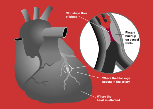
Definition
Acute Coronary Syndrome (ACS) is a spectrum of clinical presentations that arise from a sudden, reduced blood flow to the heart muscle. It encompasses unstable angina, non-ST-elevation myocardial infarction (NSTEMI), and ST-elevation myocardial infarction (STEMI).
Myocardial Infarction (MI), commonly known as a heart attack, is the irreversible death of heart muscle tissue due to prolonged ischemia (lack of oxygen).
Anatomy and physiology you need
The coronary arteries are the blood vessels that supply oxygenated blood to the heart muscle (myocardium). The two main coronary arteries are the left and right coronary arteries, which branch into smaller vessels. The left main coronary artery branches into the left anterior descending (LAD) artery and the circumflex (LCx) artery. The right coronary artery (RCA) typically supplies the inferior and posterior walls of the left ventricle and the right ventricle.
The myocardium is a muscular organ that pumps blood throughout the body. Its function is dependent on a continuous supply of oxygen and nutrients delivered by the coronary arteries. Ischemia occurs when this supply is insufficient to meet the metabolic demands of the heart muscle. Prolonged and severe ischemia leads to cellular injury and necrosis (infarction).
Causes and risk factors
The primary cause of ACS and MI is atherosclerosis, a progressive disease characterized by the buildup of plaque (fatty deposits, cholesterol, calcium, and other substances) within the coronary arteries. This plaque can narrow the arteries, restricting blood flow, or it can rupture, leading to thrombus (blood clot) formation.
Risk Factors in the Kenyan Setting:
- Modifiable:
- Hypertension: High blood pressure damages arterial walls, promoting atherosclerosis.
- Dyslipidaemia: Abnormal levels of cholesterol and triglycerides contribute to plaque formation.
- Diabetes Mellitus: A significant risk factor, often associated with accelerated atherosclerosis and microvascular disease.
- Obesity: Contributes to hypertension, dyslipidaemia, and diabetes.
- Smoking: Damages blood vessels and promotes clot formation.
- Physical Inactivity: Contributes to obesity, hypertension, and dyslipidaemia.
- Unhealthy Diet: High intake of saturated fats, trans fats, and processed foods.
- Non-Modifiable:
- Age: Risk increases with age.
- Family History: A history of premature coronary artery disease.
- Male Sex: Generally higher risk than pre-menopausal women.
- Specific to the Kenyan/Tropical Setting:
- HIV/AIDS: Chronic inflammation associated with HIV infection can accelerate atherosclerosis. Some antiretroviral therapies (ART) may also have metabolic side effects.
- Tuberculosis (TB): Chronic inflammation from TB can contribute to systemic atherosclerosis.
- Malaria: While not a direct cause of atherosclerosis, severe or recurrent malaria can lead to cardiac complications and stress on the cardiovascular system.
- Rheumatic Heart Disease: While primarily affecting heart valves, chronic inflammation and damage can indirectly impact myocardial health and increase cardiovascular risk.
- Sickle Cell Disease: Chronic haemolytic anaemia and vaso-occlusive crises can lead to increased myocardial oxygen demand, microvascular disease, and potentially contribute to cardiac dysfunction and increased risk of events.
- Other Infections/Inflammatory Conditions: Chronic inflammatory states can promote atherosclerosis.
Pathophysiology
Atherosclerotic Plaque Formation ↓ Plaque Rupture/Erosion ↓ Exposure of Thrombogenic Material (e.g., collagen) ↓ Platelet Adhesion and Activation ↓ Thrombus Formation (Blood Clot) ↓ Coronary Artery Occlusion (Partial or Complete) ↓ Myocardial Ischemia (Reduced Blood Flow) ↓ Myocardial Injury (Reversible if flow restored) ↓ Myocardial Infarction (Irreversible Necrosis if ischemia prolonged)
Clinical features
History questions that matter
- Character and site: Retrosternal pressure, heaviness, squeezing, or crushing pain radiating to the left arm, neck, jaw, or epigastrium.
- Onset and duration: Sudden onset, typically lasting greater than twenty minutes, often unprovoked or persisting at rest.
- Associated autonomic symptoms: Diaphoresis, nausea, vomiting, dizziness, and a sense of impending doom (angor animi).
- Anginal equivalents: Dyspnoea alone, unexplained fatigue, syncope, or isolated epigastric discomfort, common in women, elderly patients, and individuals with diabetes.
- Risk factors and background: Inquire about tobacco use, diabetes, hypertension, dyslipidaemia, family history, chronic kidney disease, as well as antiretroviral therapy for HIV, active tuberculosis treatment, or recreational substance use (such as cocaine, khat, or amphetamines).
Physical examination findings and pathophysiology
- General distress and pallor: Patient is pale, restless, and diaphoretic due to intense sympathetic nervous system discharge triggered by acute ischaemic stress and pain.
- Tachycardia or bradycardia: Tachycardia reflects compensatory sympathetic surge or cardiogenic shock. Bradycardia and heart blocks occur predominantly with inferior infarctions due to heightened vagal tone (Bezold-Jarisch reflex) or ischaemia to the sinoatrial/atrioventricular nodes (right coronary artery territory).
- Blood pressure variations: Hypertension arises from sympathetic drive. Hypotension indicates extensive left ventricular failure, right ventricular infarction, or mechanical rupture.
- Third heart sound ($S_3$): Caused by sudden deceleration of blood entering a non-compliant, failing, and dilated left ventricle during early diastole.
- Fourth heart sound ($S_4$): Reflects forceful atrial contraction against an acutely stiffened, non-compliant ischaemic ventricle during late diastole.
- New systolic murmur: Usually an apical pansystolic murmur radiating to the axilla, signifying acute mitral regurgitation secondary to papillary muscle ischaemia or rupture; a harsh pansystolic murmur at the left lower sternal edge suggests an acquired ventricular septal defect.
- Raised jugular venous pressure (JVP): Clear lung fields with elevated JVP and hypotension point directly to right ventricular infarction; elevated JVP with crackles indicates left-sided biventricular failure.
- Basal pulmonary crackles: Caused by elevated left ventricular end-diastolic pressure transmitting backward into pulmonary capillaries, leading to transudation of fluid into the alveoli (pulmonary oedema).
- Peripheral perfusion: Cool, clammy extremities with prolonged capillary refill time suggest falling cardiac output and cardiogenic shock.
Differential diagnosis
| Condition | What points to it | What separates it from ACS |
|---|---|---|
| Aortic dissection | Tearing pain radiating to back | Unequal pulses, normal troponin, widened mediastinum |
| Pulmonary embolism | Sudden dyspnoea, pleuritic pain | Tachycardia, S1Q3T3 on ECG, elevated D-dimer, RV strain |
| Acute pericarditis | Pleuritic pain, relieved leaning forward | Pericardial friction rub, PR depression, diffuse ST elevation |
| Tension pneumothorax | Sudden unilateral pleuritic pain | Hyper-resonance, absent breath sounds, tracheal deviation |
| Oesophageal spasm/rupture | Retrosternal pain after vomiting or food | Pain linked to swallowing, mediastinal air, normal biomarkers |
| Peptic ulcer / Pancreatitis | Epigastric pain, relation to meals | Abdominal tenderness, elevated amylase/lipase, normal ECG |
Investigations
| Test | What it answers | What a result means |
|---|---|---|
| 12-lead ECG | Presence of ischaemia, injury, or necrosis | ST elevation/LBBB indicates transmural MI; ST depression/T-inversion indicates non-transmural ischaemia |
| Cardiac troponin (I or T) | Evidence of acute myocardial necrosis | Rise and/or fall above 99th percentile confirms myocardial injury |
| Full blood count | Presence of anaemia, infection, or reactive stress | Severe anaemia precipitates type 2 MI; leukocytosis reflects acute inflammatory response |
| Urea, creatinine, electrolytes | Renal function and baseline potassium | Acute kidney injury, cardiorenal syndrome, or risk of arrhythmias from potassium imbalance |
| Bedside echocardiogram | Regional wall motion abnormalities and mechanics | Hypokinesia/akinesia marks ischaemic territory; detects acute VSD, free-wall rupture, or acute MR |
| Chest radiograph | Alternative diagnoses and pulmonary congestion | Identifies cardiogenic pulmonary oedema, widened mediastinum, or pneumothorax |
| Point-of-care blood glucose / HbA1c | Hyperglycaemia or underlying diabetes | Stress hyperglycaemia indicates poorer prognosis; identifies undiagnosed diabetes |
Diagnosis and severity
Diagnostic criteria
The Fourth Universal Definition of Myocardial Infarction requires the detection of a rise and/or fall of cardiac troponin values with at least one value above the 99th percentile upper reference limit, combined with at least one of the following:
- Symptoms of acute myocardial ischaemia.
- New ischaemic ECG changes (new ST-segment elevation, ST-segment depression, or T-wave inversion).
- Development of pathological Q waves on ECG.
- Imaging evidence of new loss of viable myocardium or new regional wall motion abnormality in a pattern consistent with an ischaemic aetiology.
- Identification of an intracoronary thrombus by angiography or autopsy.
Classification categories:
- STEMI: Clinical ischaemic symptoms with persistent (gt;20$ minutes) ST-segment elevation in two contiguous leads ($\ge 2.5\text{ mm}$ in men lt;40$ years, $\ge 2.0\text{ mm}$ in men $\ge 40$ years, or $\ge 1.5\text{ mm}$ in women in leads $V_2-V_3$; $\ge 1.0\text{ mm}$ in other leads) or new, presumed-new left bundle branch block.
- NSTE-ACS: Clinical symptoms without persistent ST elevation. Divided into NSTEMI (elevated cardiac troponin levels confirming necrosis) and Unstable Angina (normal troponin levels with transient or absent ECG changes).
Risk and severity stratification
Stratification identifies patients at high risk of rapid decompensation and death:
- Killip Classification (Post-infarct heart failure):
- Class I: No clinical signs of heart failure.
- Class II: Crackles over lower half of lung fields, $S_3$ gallop, or elevated JVP.
- Class III: Overt pulmonary oedema (crackles throughout lung fields).
- Class IV: Cardiogenic shock (systolic BP lt;90\text{ mmHg}$ with peripheral hypoperfusion).
- Clinical risk scores (GRACE and TIMI): Integrate age, heart rate, systolic blood pressure, serum creatinine, Killip class, initial cardiac arrest, ST-segment deviations, and elevated biomarkers to calculate in-hospital and 6-month mortality, directing the urgency of invasive management.
Management
Immediate Stabilisation
- Triage and positioning: Place patient on strict bed rest in a semi-Fowler position to reduce venous return and myocardial oxygen demand. Attach a continuous cardiac monitor, non-invasive blood pressure cuff, and pulse oximeter.
- Oxygen therapy: Administer supplemental oxygen only if peripheral saturation is below threshold to avoid hyperoxia-induced coronary vasoconstriction and increased reperfusion injury.
- Antiplatelet loading: Administer chewed soluble aspirin immediately to block thromboxane A2 production, combined with a P2Y12 receptor inhibitor (clopidogrel, ticagrelor, or prasugrel) to prevent further platelet aggregation and thrombus extension.
- Pain and vasodilation: Sublingual or intravenous nitrates relieve ischemic chest pain by dilating venous capacitance vessels (reducing preload) and coronary collateral channels. Intravenous opioids (e.g., morphine) reduce sympathetic drive, anxiety, and pain.
- Anticoagulation: Initiate parenteral anticoagulation using unfractionated heparin, low-molecular-weight heparin (enoxaparin), or fondaparinux to prevent ongoing thrombin generation.
Definitive Management
| ACS Type | Primary Reperfusion Strategy | Alternative Reperfusion Strategy |
|---|---|---|
| STEMI | Primary Percutaneous Coronary Intervention (PCI) | Intravenous fibrinolysis |
| High-risk NSTEMI/UA | Early invasive coronary angiography (within 24 hours) | Medical stabilisation, delayed invasive strategy |
| Low-risk NSTEMI/UA | Selective invasive strategy based on ischemia testing | Medical therapy with risk stratification |
- STEMI reperfusion: Primary PCI is the preferred modality if achievable within standard transfer windows. If delayed beyond target windows, administer intravenous fibrinolytic therapy (e.g., tenecteplase, alteplase, or streptokinase) unless absolute contraindications exist. Following fibrinolysis, arrange urgent transfer for rescue PCI (if failed reperfusion) or routine post-thrombolysis angiography.
- NSTEMI/Unstable Angina: Reperfusion with fibrinolytics is contraindicated. Manage with anti-ischemic medical therapy, parenteral anticoagulation, dual antiplatelets, and risk-stratify for urgent versus elective coronary angiography.
- In-hospital secondary prevention:
- Oral beta-blockers (e.g., carvedilol, metoprolol) to reduce heart rate, myocardial contractility, and ventricular arrhythmias.
- High-intensity statins (e.g., atorvastatin, rosuvastatin) for plaque stabilisation and lipid lowering, regardless of baseline lipid levels.
- Angiotensin-converting enzyme (ACE) inhibitors or angiotensin receptor blockers (ARBs) to prevent adverse ventricular remodeling, particularly in anterior infarction or reduced ejection fraction.
- Mineralocorticoid receptor antagonists (e.g., spironolactone) if ejection fraction is significantly depressed with heart failure signs.
Monitoring, Follow-up, and Safety
- Continuous ECG monitoring: Watch for ventricular tachyarrhythmias, heart blocks, and ST-segment resolution or re-elevation.
- Monitoring laboratory values: Trend troponin, renal function, electrolytes (keep potassium and magnesium optimal to prevent ventricular irritability), and full blood count (screen for post-infarct anemia or bleeding).
- Contraindications and safety alerts:
- Avoid nitrates in inferior STEMI with suspected right ventricular infarction, significant bradycardia, marked hypotension, or recent use of phosphodiesterase-5 inhibitors due to catastrophic preload collapse.
- Absolute contraindications to fibrinolytics include previous hemorrhagic stroke, ischemic stroke within six months, known central nervous system neoplasm or vascular malformation, recent major trauma/surgery, active internal bleeding, or suspected aortic dissection.
Complications and Prognosis
- Electrical complications: Ventricular tachycardia, ventricular fibrillation, sinus bradycardia, and high-degree atrioventricular block (common in RCA/inferior infarctions).
- Mechanical complications (typically days 3–7 post-MI):
- Papillary muscle rupture producing severe acute mitral regurgitation and sudden flash pulmonary edema.
- Ventricular septal rupture causing a harsh pansystolic murmur, biventricular failure, and left-to-right shunt.
- Left ventricular free wall rupture resulting in acute hemopericardium, cardiac tamponade, electromechanical dissociation, and death.
- Left ventricular aneurysm formation causing persistent ST-segment elevation, chronic heart failure, and systemic thromboembolism.
- Inflammatory complications: Early infarct-associated pericarditis and late autoimmune post-cardiac injury syndrome (Dressler syndrome).
- Prognosis: Determined primarily by left ventricular ejection fraction, patient age, infarct size, presence of cardiogenic shock, and timeliness of successful reperfusion.
High-Yield Exam Points
- The right ventricular STEMI trap: An inferior STEMI (leads II, III, aVF) with hypotension and clear lung fields points to right ventricular involvement; giving nitrates or morphine will drop preload and trigger profound refractory shock. Obtain right-sided leads (V4R).
- Normal initial ECG: A normal baseline ECG does not rule out unstable angina or early evolving myocardial infarction; serial tracings and troponins are mandatory.
- Fibrinolysis in NSTEMI: Never give fibrinolytic agents in NSTEMI or unstable angina; trials show no mortality benefit and an increased risk of fatal hemorrhage.
- The "silent" presentation: Elderly patients, females, and patients with diabetic autonomic neuropathy frequently present without typical retrosternal pain, manifesting instead with isolated breathlessness, syncope, delirium, or vomiting.
- Murmur after MI: A new harsh pansystolic murmur appearing days after an infarction signifies either acute ventricular septal rupture or acute mitral regurgitation from papillary muscle disruption; both require emergent echocardiography and surgical salvage.
Clinical Case
A 58-year-old male with long-standing diabetes presents to the casualty department with three hours of severe retrosternal squeezing pain radiating to his jaw, accompanied by profuse diaphoresis and vomiting. Blood pressure is normal, heart rate is regular, and chest auscultation is clear. ECG demonstrates 3 mm ST-segment elevation in leads V1 through V4.
Questions and Model Answers
- What is the definitive diagnosis and the primary culprit coronary artery?
- Acute anterior STEMI secondary to acute occlusion of the left anterior descending (LAD) coronary artery.
- The nearest PCI-capable catheterisation laboratory is five hours away. What is the immediate reperfusion strategy of choice, assuming no contraindications?
- Intravenous fibrinolysis (e.g., tenecteplase or streptokinase) administered immediately alongside dual antiplatelet therapy and anticoagulation, followed by arrangements for transfer to a PCI facility.
- On day four, he develops sudden severe dyspnea and hypotension. Examination reveals a new, loud apical pansystolic murmur and bilateral lung crackles. What complication has occurred?
- Acute severe mitral regurgitation secondary to ischemic rupture of the papillary muscle.
Practice Questions
Question 1
A 62-year-old male is admitted with an acute inferior wall STEMI. His blood pressure drops significantly after receiving a single dose of sublingual nitroglycerin. Auscultation reveals clear lung bases. What should be done next to clarify the diagnosis?
A. Perform immediate urgent pericardiocentesis B. Obtain a 12-lead ECG with right-sided precordial leads (V3R–V6R) C. Administer an intravenous loop diuretic D. Escalate intravenous opioid analgesia
Answer: B. Obtain a 12-lead ECG with right-sided precordial leads (V3R–V6R) Explanation: Inferior myocardial infarctions frequently extend into the right ventricle, creating preload dependence where nitrates cause profound hypotension; right-sided leads detect ST elevation in V4R.
Question 2
A 50-year-old female presents with rest angina and dynamic T-wave inversions in leads V4–V6. Her high-sensitivity cardiac troponin is elevated. Which medication is contraindicated in this patient's definitive management?
A. Aspirin B. Low-molecular-weight heparin C. Tenecteplase D. High-intensity atorvastatin
Answer: C. Tenecteplase Explanation: Fibrinolytic therapy is strictly contraindicated in NSTEMI because it provides no clinical benefit and increases mortality and major bleeding risks compared to antiplatelet and antithrombotic therapy.
Question 3
A 68-year-old man on day five following a transmural myocardial infarction suddenly collapses. The monitor shows sinus rhythm at a rate of 90 beats per minute, but the patient is pulseless, unresponsive, and heart sounds are inaudible. What mechanical complication has most likely occurred?
A. Left ventricular free wall rupture B. Papillary muscle rupture C. Ventricular septal rupture D. Left ventricular apical pseudoaneurysm
Answer: A. Left ventricular free wall rupture Explanation: Free wall rupture leads to rapid hemopericardium, acute cardiac tamponade, and pulseless electrical activity (electromechanical dissociation).
Question 4
A 45-year-old woman with retrosternal chest pain has an ECG showing ST elevation in leads II, III, and aVF with reciprocal depression in I and aVL. The facility cannot provide PCI within 120 minutes. What absolute contraindication must be ruled out before administering intravenous streptokinase?
A. Poorly controlled chronic hypertension without end-organ damage B. History of an ischemic stroke three years ago C. Previous hemorrhagic stroke at any time D. Concurrent use of aspirin and clopidogrel
Answer: C. Previous hemorrhagic stroke at any time Explanation: A prior history of intracerebral hemorrhage is an absolute contraindication to fibrinolytic therapy due to the high risk of recurrent fatal intracranial bleeding.
Question 5
Why are beta-blockers routinely prescribed early during the management of an uncomplicated acute coronary syndrome?
A. They dissolve microvascular coronary thrombi B. They reduce myocardial oxygen demand and suppress ventricular tachyarrhythmias C. They prevent progressive left ventricular dilation via direct inhibition of aldosterone D. They selectively dilate the epicardial coronary vessels
Answer: B. They reduce myocardial oxygen demand and suppress ventricular tachyarrhythmias Explanation: Beta-blockers decrease heart rate, systemic blood pressure, and myocardial contractility, conserving oxygen and reducing the incidence of malignant ventricular arrhythmias.
Drafted with AI assistance from standard references and reviewed for structure. Check details against your lecture notes and the Kenya MoH guideline.