By the end of this study note, the student will be able to define, classify, identify risk factors for, and explain the pathophysiologic mechanisms of cardiomyopathies; always verify drug selections, doses, and clinical pathways against the Kenya Ministry of Health guidelines and institutional formularies.
Definition
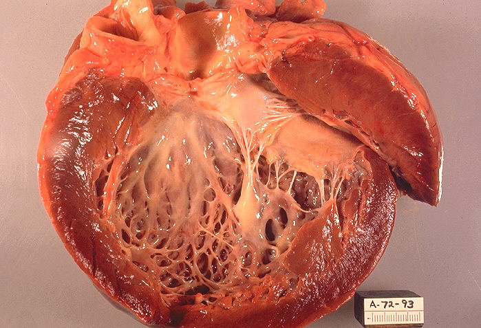
Cardiomyopathies are a heterogeneous group of myocardial diseases characterized by structural and functional abnormalities of the ventricular myocardium that are not secondary to coronary artery disease, systemic hypertension, congenital heart disease, or primary valvular disease. They are classified phenotypically into:
- Dilated Cardiomyopathy (DCM): Ventricular dilation accompanied by impaired systolic contraction (reduced left ventricular ejection fraction [LVEF < 40%]).
- Hypertrophic Cardiomyopathy (HCM): Inappropriate, unexplained myocardial hypertrophy (wall thickness $\ge 15\text{ mm}$ in adults, or $\ge 13\text{ mm}$ with a family history) occurring in the absence of abnormal loading conditions.
- Restrictive Cardiomyopathy (RCM): Impaired ventricular filling and increased myocardial stiffness with normal or near-normal wall thickness, preserved systolic function, and dilated biatrial chambers.
- Arrhythmogenic Right Ventricular Cardiomyopathy (ARVC): Progressive fibrofatty replacement of the right (and occasionally left) ventricular myocardium, predisposing to lethal ventricular arrhythmias.
Anatomy and Physiology You Need
- Myocardial Architecture: The ventricular myocardium consists of helical arrays of cardiac myocytes bound by a collagen-rich extracellular matrix. Intercalated discs couple cells mechanically via desmosomes and electrically through gap junctions (connexin-43), ensuring synchronized mechanical systole.
- Excitation-Contraction Coupling: Action potential propagation triggers calcium influx via L-type calcium channels, initiating calcium-induced calcium release from the sarcoplasmic reticulum through ryanodine receptors. Calcium binds troponin C, releasing tropomyosin inhibition to allow actin-myosin cross-bridge cycling. Active cross-bridge detachment and calcium re-uptake by SERCA2a govern lusitropy (diastolic relaxation).
- Frank-Starling Relationship: Increasing end-diastolic sarcomere stretch elevates calcium sensitivity, augmenting stroke volume. Excessive dilation or progressive matrix disarray disrupts sarcomere alignment, shifting the operating curve downward and causing forward low-output failure and elevated filling pressures.
- Coronary Perfusion Dynamics: Left ventricular subendocardial perfusion occurs almost exclusively during diastole. When diastolic pressures rise or ventricular wall thickness increases without proportional capillary angiogenesis (as in HCM), the myocardial oxygen supply-demand balance fails, inducing ischemia without epicardial coronary occlusion.
Causes and Risk Factors
| Phenotype | Primary / Genetic Causes | Tropical, Infectious & Acquired Causes (Regional Focus) |
|---|---|---|
| DCM | Titin (TTN), lamin A/C (LMNA) mutations | Post-viral myocarditis, advanced HIV infection, peripartum state, chronic alcohol use |
| HCM | Sarcomeric mutations (MYH7, MYBPC3) | Storage disorders (Fabry disease, glycogen storage) |
| RCM | Sarcomeric mutations, familial amyloidosis | Endomyocardial fibrosis, cardiac tuberculosis, untreated sickle cell disease (iron overload) |
| ARVC | Desmosomal gene mutations (PKP2, DSP) | Chagas disease (rare/imported), inflammatory mimickers |
- Infectious & Immune-mediated: Advanced HIV causes direct myocyte toxicity, cytokine-driven apoptosis, or secondary myocarditis via opportunistic pathogens (e.g., cytomegalovirus, toxoplasmosis). Tuberculosis leads to myocardial tuberculomas or chronic constrictive-restrictive myopericarditis. Enteroviruses (Coxsackievirus B) and arboviruses cause post-infectious inflammatory DCM.
- Infiltrative & Interstitial: Endomyocardial fibrosis (EMF)—endemic to warm, humid tropical belts—causes apical obliteration and severe restriction via subendocardial fibrous tissue deposition. Secondary hemochromatosis from repeated blood transfusions in severe sickle cell anemia induces myocardial iron overload, causing secondary RCM transitioning to DCM.
- Metabolic & Nutritional: Severe micronutrient deficits (thiamine deficiency causing wet beriberi, selenium deficiency) and severe chronic anemia trigger high-output states that eventually cause secondary myocardial exhaustion and biventricular failure.
- Peripartum Cardiomyopathy (PPCM): Idiopathic systolic failure presenting between the final month of pregnancy and five months postpartum; risk factors include multiparity, pre-eclampsia, advanced maternal age, and twin gestations, driven by vascular-damaging prolactin cleavage fragments ($16\text{-kDa}$ prolactin).
Pathophysiology
Dilated Cardiomyopathy (DCM)
Genetic defect, viral infection, or toxin $\rightarrow$ Progressive myocyte necrosis and interstitial fibrosis $\rightarrow$ Sarcomere slippage with eccentric ventricular remodeling $\rightarrow$ Left ventricular chamber enlargement with thinned walls $\rightarrow$ Systolic pump failure (depressed LVEF) $\rightarrow$ Secondary functional mitral regurgitation due to papillary muscle displacement $\rightarrow$ Chronic elevation of left atrial and pulmonary venous pressures $\rightarrow$ Biventricular congestion and forward cardiogenic shock.
Hypertrophic Cardiomyopathy (HCM)
Mutations in sarcomeric contractile proteins $\rightarrow$ Disorganized, chaotic myofibrillar architecture (myocyte disarray) $\rightarrow$ Asymmetric septal hypertrophy $\rightarrow$ Ventricular compliance decreases, driving severe diastolic dysfunction $\rightarrow$ Systolic anterior motion (SAM) of the anterior mitral valve leaflet toward the hypertrophied septum $\rightarrow$ Dynamic left ventricular outflow tract (LVOT) obstruction $\rightarrow$ Increased intraventricular pressures, microvascular ischemia, and malignant re-entrant ventricular tachyarrhythmias.
Restrictive Cardiomyopathy (RCM)
Endomyocardial fibrosis, granulomatous deposition, or iron overload $\rightarrow$ Massive accumulation of fibrotic tissue or abnormal proteins within the myocardial interstitium $\rightarrow$ Rigid, non-compliant ventricular myocardium $\rightarrow$ Severe impairment of ventricular diastolic filling with preserved early systolic ejection $\rightarrow$ Rapid, precipitous rise in early-to-mid diastolic intraventricular pressures $\rightarrow$ Markedly elevated systemic and pulmonary venous pressures with massive biatrial enlargement.
Clinical features
History questions that matter
- Onset and progression of dyspnea: Inquire about NYHA functional class, orthopnea, and paroxysmal nocturnal dyspnea (PND) to grade left ventricular failure and pulmonary venous congestion.
- Chest pain and exertional syncope: Critical in hypertrophic cardiomyopathy (HCM) where exertional syncope signifies dynamic left ventricular outflow tract (LVOT) obstruction or ventricular arrhythmias.
- Systemic and tropical risk factors: Recent delivery (peripartum cardiomyopathy, typically within 5 months postpartum); heavy alcohol intake; chemotherapy exposure (e.g., doxorubicin).
- Infections and chronic illness: Known HIV infection, current or past tuberculosis (TB), chronic inflammatory states, or a history of nutritional deprivation (thiamine deficiency/wet beriberi).
- Family history: Inquire specifically about unexplained premature death or sudden cardiac arrest in first-degree relatives under 40 years (familial dilated cardiomyopathy [DCM], HCM, or arrhythmogenic right ventricular cardiomyopathy [ARVC]).
Examination findings and why each occurs
- Elevated Jugular Venous Pressure (JVP): Reflects high right atrial pressure secondary to elevated right ventricular end-diastolic pressure or biventricular failure. Look for a prominent v wave if functional tricuspid regurgitation is present, or a positive Kussmaul sign (paradoxical rise in JVP on inspiration) typical of restrictive cardiomyopathy (RCM).
- Displaced, diffuse apex beat: Shifted laterally and inferiorly beyond the 5th intercostal space, midclavicular line in DCM, reflecting marked left ventricular dilatation and eccentric remodeling.
- Double or triple apical impulse: Pathognomonic for HCM; represents a palpable atrial kick (forceful left atrial contraction against a non-compliant LV) followed by sustained ventricular systolic thrust.
- Auscultation - Heart sounds:
- Third heart sound (S3): Caused by rapid, passive ventricular filling into an already overloaded, dilated, and non-compliant LV chamber (hallmark of DCM).
- Fourth heart sound (S4): Caused by vigorous atrial systole into a stiff, hypertrophied, non-compliant ventricle (frequent in HCM and amyloid RCM; absent in atrial fibrillation).
- Murmurs:
- Holosystolic murmur at the apex (radiating to axilla): Functional mitral regurgitation resulting from annular dilatation, papillary muscle displacement, and ventricular remodeling in DCM.
- Harsh, crescendo-decrescendo systolic murmur at left sternal border: Produced by dynamic subaortic narrowing in HCM. It intensifies with maneuvers that decrease LV preload/end-diastolic volume (Valsalva strain phase, standing) and softens with maneuvers that increase preload or afterload (squatting, passive leg raise).
- Pulmonary crackles (crepitations): Bibasilar, fine, end-inspiratory crackles caused by transudative alveolar fluid secondary to raised pulmonary capillary wedge pressure.
- Peripheral edema, tender hepatomegaly, and ascites: Result from systemic venous congestion due to right-sided heart failure.
Differential diagnosis
| Condition | What points to it | What separates it from this one |
|---|---|---|
| Hypertensive heart disease | Longstanding elevated blood pressure readings | Symmetric hypertrophy; clears when load drops |
| Severe aortic stenosis | Systolic ejection murmur radiating to carotids | Fixed valvular calcification/narrowing on echo; parvus et tardus pulse |
| Pericardial constriction | Kussmaul sign, pericardial knock, high JVP | Pericardial thickening/calcification; normal myocardial tissue Doppler |
| Rheumatic heart disease | History of carditis, localized primary organic murmurs | Primary valvular destruction/commissural fusion rather than functional regurgitation |
| Cor pulmonale | Chronic lung disease, isolated right-sided failure | Absence of primary left ventricular remodeling; pulmonary pathology |
| Ischemic cardiomyopathy | Prior myocardial infarction, focal angina pectoris | Regional wall motion abnormalities matching single coronary artery territory |
| Cardiac amyloidosis | Low ECG voltage despite thick ventricular walls | Granular/sparkling myocardial texture with abnormal strain imaging |
Investigations
| Test | What it answers | What a result means |
|---|---|---|
| 12-lead ECG | Rhythm, conduction defects, chamber hypertrophy | Low voltage (amyloid/pericardial fluid); massive voltage with deep lateral T-inversions (HCM); LBBB (dyssynchrony) |
| Chest X-ray | Cardiac chamber profile and pulmonary hemodynamics | Cardiothoracic ratio >0.55 in DCM; upper lobe diversion and Kerley B lines indicate pulmonary venous congestion |
| Transthoracic echocardiogram | Definitive structural phenotype and ventricular function | Dilated chambers with EF <40% (DCM); asymmetric septal hypertrophy >15 mm (HCM); biatrial enlargement with normal cavity size (RCM) |
| Full blood count & ESR/CRP | Intercurrent infection, anemia, or systemic inflammation | Severe anemia causing high-output failure; raised markers suggesting myocarditis or active systemic disease |
| Serum electrolytes, urea, creatinine | Baseline renal status, cardiorenal syndrome | Hyponatremia indicates severe neurohormonal activation; monitors safe initiation of renin-angiotensin inhibitors |
| HIV serology and CD4 count | Secondary etiology common in local setting | Identifies HIV-associated cardiomyopathy or myocarditis driven by viral reservoir or opportunistic processes |
| Serum ferritin and iron studies | Underlying secondary metabolic cause | Elevated saturation suggests hemochromatosis causing secondary DCM or RCM |
Diagnosis and severity
Diagnostic confirmation
Diagnosis requires clinical suspicion substantiated by objective transthoracic echocardiography, after excluding coronary artery disease, abnormal loading conditions (uncontrolled hypertension, primary valvular disease), and congenital shunts adequate to cause the phenotype.
- DCM: Left ventricular ejection fraction (LVEF) <40% (or fractional shortening <25%) combined with left ventricular end-diastolic dimension (LVEDD) >112% of predicted normal values.
- HCM: Unexplained maximal left ventricular wall thickness ≥15 mm in any myocardial segment (or ≥13 mm in a patient with a confirmed first-degree relative with HCM).
- RCM: Restrictive filling pattern (mitral inflow E/A ratio >2, shortened E-deceleration time <150 ms, reduced tissue Doppler early diastolic velocities [e']) with normal or near-normal LV volumes and preserved LVEF.
Severity assessment
- Functional severity: Classified via the New York Heart Association (NYHA) system (Class I: asymptomatic ordinary activity; Class II: slight limitation; Class III: marked limitation on minimal activity; Class IV: symptoms at rest).
- Hemodynamic and electrical severity markers:
- Persistent resting tachycardia, hypotension (systolic blood pressure <90 mmHg), or refractory fluid overload.
- Severe systolic impairment (LVEF <30%).
- Elevated systemic biomarkers (cardiac troponins signifying continuing myocyte injury; elevated natriuretic peptides reflecting elevated wall tension).
- Presence of complex ventricular arrhythmias, non-sustained ventricular tachycardia (NSVT) on telemetry/Holter, or advanced conduction blocks.
- For HCM: LVOT resting or provocable peak gradient ≥50 mmHg, personal history of unexplained syncope, or massive hypertrophy (wall thickness ≥30 mm).
Management
Immediate Stabilisation
- Airway and Breathing: Sit the patient upright. Administer high-flow supplemental oxygen via face mask if oxygen saturation falls below 90% or if the patient exhibits respiratory exhaustion. Initiate non-invasive positive pressure ventilation (CPAP or BiPAP) for acute cardiogenic pulmonary oedema to reduce preload and afterload while recruiting collapsed alveoli.
- Circulation: Gain intravenous access. In acute dilated cardiomyopathy (DCM) with volume overload, give an intravenous loop diuretic (furosemide) to induce rapid venodilatation and natriuresis.
- Vasodilators: If systolic blood pressure remains adequate, start an intravenous nitrate infusion (glyceryl trinitrate) to unload the left ventricle by reducing systemic vascular resistance and pulmonary capillary wedge pressure.
- Cardiogenic Shock: If perfusion fails (cold peripheries, altered mentation, oliguria), avoid vasodilators and aggressive fluid boluses. Start an inotrope (dobutamine) or an inodilator (milrinone) to restore forward flow, adding a vasopressor (noradrenaline) if profound hypotension coexists.
- Tachyarrhythmias: Unstable ventricular tachycardia or rapid atrial fibrillation demands immediate synchronised electrical cardioversion.
Definitive Treatment
| Cardiomyopathy Type | Pharmacotherapy | Interventions & Devices |
|---|---|---|
| Dilated (DCM) | ACEi/ARNI, beta-blocker, MRA, SGLT2i | ICD for sudden death risk; CRT-P/D |
| Hypertrophic (HCM) | Non-vasodilating beta-blockers, verapamil | Septal myectomy, alcohol septal ablation, ICD |
| Restrictive (RCM) | Cautious loop diuretics; treat primary cause | Endomyocardial resection; cardiac transplant |
| Peripartum (PPCM) | Standard HFrEF therapy; bromocriptine | Anticoagulation while EF severely reduced |
- Dilated Cardiomyopathy: Once euvolaemic, establish the four foundational pillars of heart failure therapy: an ACE inhibitor, ARB, or ARNI (sacubitril-valsartan); an evidence-based beta-blocker (carvedilol, bisoprolol, or sustained-release metoprolol); a mineralocorticoid receptor antagonist (spironolactone); and an SGLT2 inhibitor (dapagliflozin or empagliflozin). Add oral loop diuretics as needed for symptom control. Address underlying tropical triggers: optimise antiretroviral therapy for HIV-associated cardiomyopathy, initiate full-course antituberculous therapy for TB myocarditis, and correct severe chronic anaemia.
- Hypertrophic Cardiomyopathy: Beta-blockers (propranolol, metoprolol, atenolol) form first-line therapy to slow the heart rate, prolong diastolic filling, and diminish dynamic left ventricular outflow tract (LVOT) obstruction. Use non-dihydropyridine calcium channel blockers (verapamil) if beta-blockers fail or are contraindicated.
- Restrictive Cardiomyopathy: Diuretics must be titrated with extreme caution; excessively lowering filling pressures precipitates severe low-output hypotension because these stiff ventricles depend on elevated preload to fill. Treat systemic disorders (e.g., iron chelation for haemochromatosis; corticosteroids/chemotherapy for amyloidosis or sarcoidosis).
- Anticoagulation: Warfarin or a direct oral anticoagulant (DOAC) is indicated in DCM with atrial fibrillation, previous thromboembolism, or demonstrable LV mural thrombus, as well as in PPCM when the ejection fraction remains below 35% due to high peripartum prothrombotic risk.
Monitoring and Safety
- Renal Function and Electrolytes: Check baseline serum creatinine, urea, and potassium before starting ACE inhibitors, ARNIs, or spironolactone. Re-evaluate at 1 to 2 weeks post-initiation and after dose escalations.
- Blood Pressure and Heart Rate: Monitor for symptomatic hypotension and bradycardia during beta-blocker up-titration. Beta-blockers must never be initiated during an acute, decompensated "wet and cold" heart failure state; the patient must be dry and stabilised first.
- Drug Contraindications in HCM: Strictly avoid pure vasodilators (dihydropyridine calcium channel blockers, nitrates, ACE inhibitors) and inotropes (digoxin) in obstructive HCM; reducing chamber size or increasing contractility worsens the dynamic LVOT gradient and triggers cardiovascular collapse.
Complications and Prognosis
- Acute Decompensated Heart Failure: Rapid pulmonary oedema precipitated by non-adherence, intercurrent infection (e.g., malaria, pneumonia), or dietary sodium loading.
- Malignant Arrhythmias: Ventricular tachycardia and ventricular fibrillation causing sudden cardiac death (highest in HCM and advanced DCM).
- Systemic and Pulmonary Thromboembolism: Mural thrombus dislodgement causes ischaemic stroke, renal infarction, or limb ischaemia; deep vein thromboses lead to pulmonary embolism.
- Functional Valvular Regurgitation: Progressive annular dilatation causes severe functional mitral or tricuspid regurgitation, accelerating biventricular failure.
- Conduction System Disease: Complete heart block secondary to infiltrative diseases (cardiac amyloidosis, sarcoidosis).
- Prognosis: PPCM carries a reasonable recovery rate if the ventricle normalises within 6 months, though subsequent pregnancy carries high recurrence risk. End-stage DCM and amyloid RCM have poor long-term outcomes without advanced device support or heart transplantation.
High-Yield Exam Points
- The Obstructive HCM Murmur Trap: The systolic murmur of obstructive HCM increases with the Valsalva strain phase and standing (decreased preload narrows the outflow tract), whereas the murmur of aortic stenosis decreases (less flow across the fixed valve).
- The "Look-Alike" RCM vs Constrictive Pericarditis: Examiners love asking how to separate them. Constrictive pericarditis features pericardial knock, pericardial calcification on chest X-ray, equalisation of end-diastolic pressures in all four cardiac chambers, and preserved tissue Doppler lateral mitral annular velocity (annulus reversus). Restrictive cardiomyopathy shows markedy elevated BNP, blunted annular tissue velocities, and thick ventricular walls.
- The Fatal Bedside Error in HCM: Prescribing nitrates or furosemide to an acutely breathless patient with unsuspected hypertrophic obstructive cardiomyopathy. Decreasing venous return precipitates catastrophic outflow tract obstruction and sudden death.
- PPCM Timing: Symptoms strictly arise in the final month of pregnancy up to 5 months postpartum without prior identifiable heart disease. Heart failure developing in the second trimester is usually unmasked pre-existing rheumatic heart disease or congenital heart disease.
- Digoxin Danger: Never give digoxin in HCM (worsens obstruction through positive inotropy) or in amyloid RCM (amyloid fibrils bind digitalis, causing fatal toxicity even at standard serum concentrations).
Clinical Case
A 28-year-old female (gravida 3, para 3) presents to the emergency department 6 weeks following an uncomplicated home delivery with progressive orthopnoea, bilateral leg swelling, and dry cough. Examination reveals a blood pressure of 100/65 mmHg, heart rate of 115 bpm, elevated jugular venous pressure (6 cm above the sternal angle), bilateral basal crackles extending halfway up the lung fields, an S3 gallop, and a soft pansystolic murmur at the apex. Echocardiography demonstrates global left ventricular hypokinesia with an ejection fraction of 28% and mild mitral regurgitation.
Questions and Model Answers
- What is the most likely diagnosis?
Peripartum cardiomyopathy (PPCM) presenting with acute decompensated heart failure with reduced ejection fraction.
- Which two non-heart failure medications must be added immediately to her treatment regimen, and why?
Anticoagulation (low-molecular-weight heparin transitioning to warfarin or DOAC) because an ejection fraction below 30% in the puerperium creates an exceptionally high risk of left ventricular mural thrombus and thromboembolism; and bromocriptine (under physician guidance, with prophylactic anticoagulation) to inhibit prolactin cleavage into cardiotoxic 16-kDa prolactin fragments.
- What crucial counselling regarding future family planning must this patient receive?
She must be counselled on the high risk of heart failure recurrence, persistent myocardial damage, and maternal mortality in subsequent pregnancies, particularly if her left ventricular ejection fraction fails to recover completely. Reliable contraception (avoiding combined oestrogen-containing oral contraceptives due to thrombosis risk) should be provided.
Practice Questions
Question 1
A 45-year-old male with long-standing, well-controlled HIV infection presents with fatigue and exertional dyspnoea. Clinical examination reveals an apical impulse displaced to the 6th intercostal space, anterior axillary line, an audible third heart sound, and an elevated JVP. Echocardiography confirms dilated cardiomyopathy with an ejection fraction of 30%. The patient is clinically euvolaemic on oral furosemide. Which of the following is the most appropriate next step in management?
A. Add verapamil to control resting tachycardia B. Stop antiretroviral therapy to eliminate drug-induced cardiotoxicity C. Initiate carvedilol at a low dose with gradual up-titration D. Commence an intravenous dobutamine infusion
Answer: C. Initiate carvedilol at a low dose with gradual up-titration Explanation: Guideline-directed medical therapy for HFrEF requires early introduction of an evidence-based beta-blocker once the patient is clinically stable and euvolaemic. Verapamil is contraindicated in reduced ejection fraction due to negative inotropy, and ART should be maintained to control HIV replication.
Question 2
A 22-year-old athlete collapses during a local football match. On arrival, he is conscious. Bedside examination reveals a harsh, crescendo-decrescendo systolic murmur heard best along the left lower sternal border. The murmur intensifies significantly when the patient performs a Valsalva manoeuvre. Which medication is strictly contraindicated?
A. Bisoprolol B. Verapamil C. Glyceryl trinitrate D. Disopyramide
Answer: C. Glyceryl trinitrate Explanation: The findings are classic for hypertrophic obstructive cardiomyopathy (HOCM). Vasodilators such as nitrates decrease venous return and systemic vascular resistance, reducing LV chamber volume and worsening dynamic LVOT obstruction, which can cause syncope or arrest.
Question 3
A 52-year-old woman with treated pulmonary tuberculosis presents with peripheral oedema, abdominal distension, and hepatomegaly. The JVP is markedly elevated and rises further during inspiration. Echocardiography shows bilateral atrial enlargement, normal ventricular wall thickness, normal left ventricular systolic function, and impaired early diastolic ventricular filling. Why is an aggressive diuresis strategy dangerous in this patient?
A. It induces immediate malignant ventricular tachyarrhythmias B. It dramatically drops preload in non-compliant ventricles, causing severe forward failure C. It provokes left ventricular outflow tract dynamic obstruction D. It induces severe secondary hyperkalaemia
Answer: B. It dramatically drops preload in non-compliant ventricles, causing severe forward failure Explanation: In restrictive cardiomyopathy, stiff, non-compliant ventricles require high filling pressures (preload) to achieve adequate end-diastolic volume. Over-diuresis precipitates a critical drop in stroke volume and profound hypotension.
Question 4
A 34-year-old woman presents 3 weeks postpartum with sudden-onset left hemiparesis. She was diagnosed with peripartum cardiomyopathy 10 days ago (ejection fraction 22%). Brain imaging confirms an acute middle cerebral artery ischaemic infarct. What pathophysiological mechanism directly accounts for this event?
A. Paradoxical embolism from deep venous thrombosis through an atrial septal defect B. Formation and dislodgement of a left ventricular mural thrombus due to severe stasis C. Accelerated postpartum intracranial atherosclerotic plaque rupture D. Endocarditis-induced septic embolisation from the mitral valve
Answer: B. Formation and dislodgement of a left ventricular mural thrombus due to severe stasis Explanation: Severe systolic dysfunction (EF < 30%) combined with the postpartum hypercoagulable state promotes stasis in the apex of the dilated left ventricle, leading to mural thrombus formation and subsequent cerebral embolisation.
Question 5
A 48-year-old man with dilated cardiomyopathy and an ejection fraction of 25% is admitted with acute, severe shortness of breath, bilateral crackles, cold extremities, and a blood pressure of 78/50 mmHg. Which choice represents the correct initial medical strategy?
A. Intravenous propranolol and oral spironolactone B. Aggressive intravenous boluses of normal saline C. Intravenous inotropic support with dobutamine and a loop diuretic D. Intravenous verapamil and sublingual isosorbide dinitrate
Answer: C. Intravenous inotropic support with dobutamine and a loop diuretic Explanation: The patient is in cardiogenic shock ("cold and wet"). An inotrope such as dobutamine is required to increase contractility and systemic perfusion, along with cautious diuresis; beta-blockers and vasodilators are contraindicated in the presence of acute hypotension and shock.
Drafted with AI assistance from standard references and reviewed for structure. Check details against your lecture notes and the Kenya MoH guideline.