Year 4 MBChB — Internal Medicine clinical skills. This guide explains the sequence, the manoeuvres, what a positive result means, and how to present findings.
1. The abdominal examination sequence
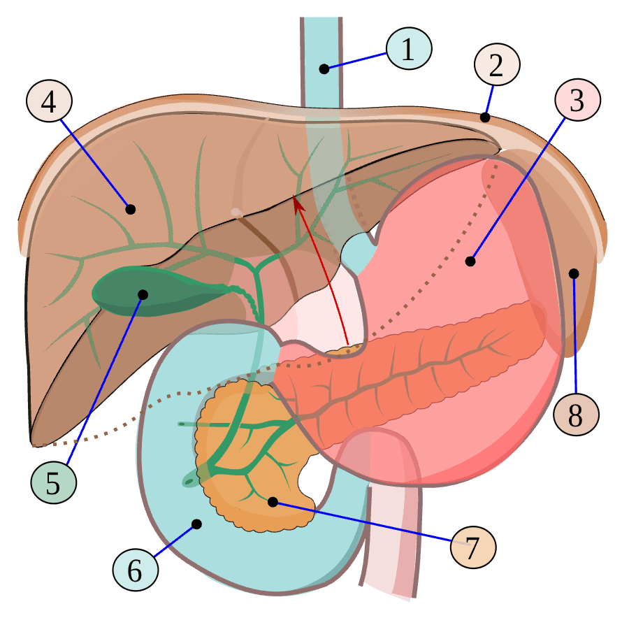
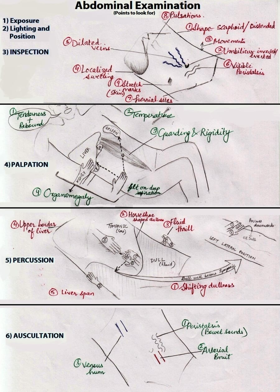
For the abdomen, use:
Inspection → Auscultation → Percussion → Palpation.
This is often remembered as IAPP. The key difference from the respiratory examination is that you auscultate the abdomen before percussion and palpation, because tapping and pressing can change bowel sounds. Some clinical teaching frameworks include special additional steps, but this is the standard bedside sequence for a routine abdominal examination.
Explain the examination, obtain consent, ask about pain, wash hands, position the patient supine with the head supported and knees slightly flexed if helpful, expose from xiphisternum to pubic region while maintaining dignity, and ensure good lighting. Warm your hands and ask where the pain is; examine painful areas last during palpation.
2. Inspection — what do you look for?
Stand at the foot or right side of the bed and inspect from different angles.
- Contour: flat, rounded, distended, scaphoid (sunken) or asymmetric.
- Movement: does the abdomen move with breathing? Is there visible peristalsis or pulsation?
- Skin: scars, striae, bruising, dilated veins, rashes, pigmentation.
- Umbilicus: central or displaced, inverted or everted, hernia, discharge.
- Masses or hernias: visible bulges; ask the patient to cough if appropriate.
- Flanks: fullness can occur with substantial ascites.
- General context: cachexia, jaundice, peripheral oedema, muscle wasting, confusion or breathlessness.
An enlarged abdomen is not automatically ascites. Causes include gas, stool, pregnancy, obesity, organ enlargement, a mass and fluid. You need to examine and investigate the patient rather than diagnose from appearance alone.
Peripheral signs of liver disease and infective endocarditis
Depending on the clinical question, inspect hands and nails for clubbing, leukonychia, palmar erythema, bruising, muscle wasting, splinter haemorrhages and other peripheral signs. Look for jaundice in the sclera and conjunctival pallor. Osler nodes are tender finger/toe-pad nodules and Janeway lesions are classically painless palm/sole lesions; they are possible signs of infective endocarditis but are neither sensitive nor specific enough to diagnose it alone.
3. Auscultation — listen before tapping or pressing
Use the diaphragm to listen for bowel sounds in several abdominal quadrants. Bowel sounds are intermittent, so listen long enough before deciding they are absent; follow your institution's examination standard. Listen for bruits over the aorta and selected renal/iliac/femoral arteries if indicated by the history and clinical context.
Do not over-interpret bowel sounds. A single short listen cannot reliably diagnose obstruction or ileus. Integrate the history, abdominal pain, vomiting, distension, bowel function and the rest of the examination.
4. Percussion — tympany and dullness
Percussion helps distinguish gas-filled bowel from solid organs, masses and fluid.
- Tympany: a drum-like note, commonly over gas-filled bowel.
- Dullness: a quieter note over solid organs, masses, stool or fluid.
- Liver dullness: helps estimate the liver's upper border and liver span.
- Shifting dullness: assesses whether dullness moves when the patient changes position, supporting the presence of free peritoneal fluid (ascites).
Percussion findings depend on technique, body habitus, bowel gas and the amount of fluid. Small-volume ascites can be missed clinically; ultrasound is more sensitive.
5. How to test for ascites
Ascites means abnormal accumulation of free fluid in the peritoneal cavity. Common causes include cirrhosis/portal hypertension, malignancy, heart failure, tuberculosis and other inflammatory or systemic conditions. The physical examination detects larger volumes better than small ones.
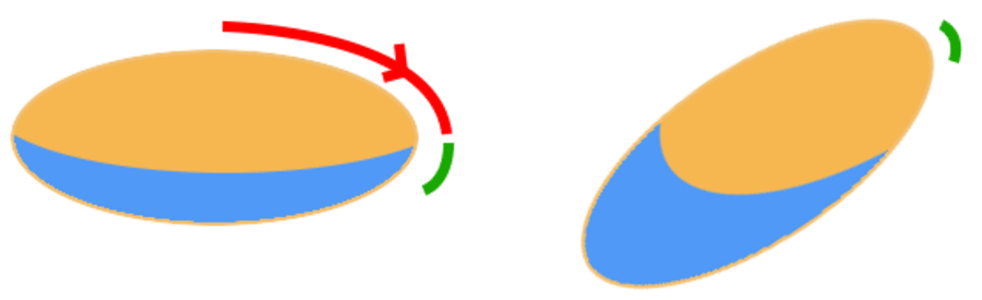
Illustration of shifting dullness. Source: Medical Abdomen teaching slide. The diagrams below describe how to perform the tests at the bedside.
A. Shifting dullness — the test you described involving turning the patient
Principle: in a supine patient with free fluid, fluid gravitates to the dependent flanks, while gas-filled bowel tends to float toward the front. When the patient turns onto their side, the fluid shifts, so the boundary between tympany and dullness changes.
Steps
- Position the patient supine.
- Percuss from the central abdomen toward one flank until the note changes from tympany to dullness.
- Keep your finger at the transition point or remember the location.
- Ask the patient to roll onto one side, if safe and tolerated. Wait briefly for fluid to move.
- Percuss again from the uppermost side toward the dependent side.
- A change in the location of the dullness boundary is a positive shifting dullness result, supporting ascites.
If the patient cannot turn, you may not be able to complete this manoeuvre reliably. Do not force the patient to move if it is unsafe or painful. Document the limitation and consider ultrasound.
B. Fluid thrill (fluid wave) — the “tap one side, feel the other” test
This is the test you were trying to remember. It looks for transmission of a wave through a large volume of fluid.
Steps
- The patient lies supine.
- Ask the patient or an assistant to place the ulnar edge of a hand firmly down the midline of the abdomen. This reduces transmission of vibration through the abdominal wall.
- Place one of your hands against one flank.
- Tap or flick the opposite flank with the other hand.
- Feel for a transmitted impulse/wave at the hand on the opposite side.
A clearly transmitted wave is a positive fluid thrill and can support large-volume ascites. It is not very sensitive for small amounts of fluid and can be falsely positive if the midline is not stabilised. The midline hand is important.
C. Fluid thrill versus shifting dullness
| Test | What you do | What a positive test suggests |
|---|---|---|
| Shifting dullness | Percuss, turn patient, percuss again | The boundary of dullness shifts with gravity |
| Fluid thrill | Stabilise midline; tap one flank and feel the other | A transmitted fluid wave, usually with larger-volume ascites |
Do not confuse a fluid thrill with a succussion splash, which is a different manoeuvre used in selected contexts to detect fluid and gas in a hollow viscus.
D. What if neither test is positive?
That does not exclude small-volume ascites. If suspicion remains—especially with chronic liver disease, oedema, raised JVP or unexplained abdominal distension—request/interpret abdominal ultrasound and assess the cause. New-onset ascites often requires diagnostic paracentesis by a trained clinician, with fluid analysis guided by local protocols.
6. Palpation — feel gently and systematically
Begin away from the painful area. Ask the patient to relax and breathe slowly.
- Light palpation: tenderness, guarding, superficial masses.
- Deep palpation: deeper masses and organ enlargement.
- Liver: palpate from the right lower abdomen upward toward the right costal margin while the patient breathes in.
- Spleen: start in the right iliac fossa and move diagonally toward the left costal margin, because a large spleen can extend down and across the abdomen.
- Kidneys and other structures: use the appropriate bimanual techniques when indicated.
- Peritonism: involuntary guarding/rigidity or pain with movement can indicate an acute abdomen and needs urgent assessment.
Do not repeatedly provoke rebound tenderness. If acute abdomen is suspected, examine carefully, escalate and follow local protocols.
7. Measuring liver span in centimetres
The liver span is the vertical distance between the upper and lower borders of liver dullness, usually assessed along the right midclavicular line.
Percussion method
- With the patient supine, percuss down from the right upper chest along the right midclavicular line to identify where resonance becomes dull (the approximate upper border).
- Starting lower down over the abdomen, percuss upward along the same line to identify where tympany becomes dull (the approximate lower border).
- Mark both levels and measure the distance between them in centimetres.
A typical adult liver span is often quoted around 6–12 cm at the right midclavicular line, but the expected range depends on sex, body size, technique and reference. Use your school's accepted range in examinations and interpret with the rest of the clinical picture.
A large span may suggest hepatomegaly, but lung hyperinflation, body habitus and technique can alter the estimate. Palpation can also help: feel for the liver edge as the patient inspires. A palpable liver edge does not automatically mean the liver is enlarged.
8. Asterixis — the “liver flap”
Asterixis is a brief, involuntary loss of sustained posture, often called a flapping tremor. It can occur in hepatic encephalopathy, but also in other metabolic/toxic encephalopathies, including uraemia and hypercapnia. It is not exclusive to liver disease.
How to test
- Ask the patient to extend both arms forward.
- Ask them to cock the wrists backwards (dorsiflex), spread the fingers and hold the position.
- Observe the hands for several seconds.
- Asterixis appears as irregular brief lapses of wrist extension, causing a flapping movement.
If the patient is confused, drowsy or has new altered consciousness, assess urgently. Do not label hepatic encephalopathy based on asterixis alone: consider glucose, medicines/toxins, infection, renal failure, carbon dioxide retention and other causes.
9. Hepatomegaly, oedema and heart failure
The liver can enlarge and become tender in right-sided heart failure because venous pressure backs up into the hepatic veins. Raised JVP, dependent oedema, ascites and a tender enlarged liver can occur together. In severe congestion, liver enlargement may be accompanied by abnormal liver blood tests.
Look for:
- Tachypnoea and breathlessness; oxygen saturation.
- Basal crackles that may suggest pulmonary congestion.
- Raised JVP.
- Bilateral pitting ankle oedema (or sacral oedema in a bedbound patient).
- Hepatomegaly and possible ascites.
- Tachycardia, cool peripheries, low blood pressure or reduced urine output in severe illness.
These signs have other causes and should be interpreted together. Suspected acute decompensated heart failure needs timely clinical assessment.
10. Pitting peripheral oedema
Press a thumb firmly for several seconds over the shin just above the ankle or over the sacrum in a bedbound patient. A persistent indentation is pitting oedema. Compare sides and document extent/severity using your institution's grading method. Causes include heart failure, renal disease, liver disease, venous insufficiency, malnutrition and some medications.
11. What to say in an OSCE
Normal abdominal examination:
“The abdomen is not distended, with no visible scars or dilated veins. Bowel sounds are present. The abdomen is soft and non-tender, with no palpable masses or organomegaly. Percussion is predominantly tympanitic, with normal liver span on percussion and no clinical evidence of ascites.”
Only state findings you actually assessed; adjust the wording to the examination.
Distended abdomen with suspected ascites:
“The abdomen is distended with flank fullness. There is shifting dullness [and/or a fluid thrill] on examination, supporting ascites. I would assess for signs of chronic liver disease and cardiac/renal causes, check vital signs and arrange abdominal ultrasound and appropriate blood tests. New-onset ascites requires clinical review and may need diagnostic paracentesis.”
Possible congestive hepatomegaly:
“There is a palpable, possibly tender liver edge, with raised JVP and bilateral pitting oedema. This raises concern for systemic venous congestion/right-sided heart failure, but I would integrate the full cardiovascular and respiratory examination and investigations.”
12. Quick recall for the exam
- Chest/respiratory: IPPA — inspection, palpation, percussion, auscultation.
- Abdomen: IAPP — inspection, auscultation, percussion, palpation.
- Shifting dullness: percuss → turn the patient → percuss again; dullness shifts with fluid.
- Fluid thrill: stabilise midline → tap one flank → feel the other.
- Liver span: identify upper and lower borders of dullness on the right midclavicular line and measure between them.
- Asterixis: arms extended, wrists dorsiflexed, fingers spread; look for brief lapses of posture.
- A distended abdomen is not automatically ascites. Clinical examination and ultrasound help confirm it and identify the cause.
Further reading
- Merck Manual Professional: Evaluation of abdominal symptoms
- NCBI Bookshelf: Clinical examination and ascites
- Shifting dullness teaching illustration
Next topic: CNS examination — mental status, cranial nerves, tone, power, reflexes, sensation, coordination, gait and meningeal signs.
13. Additional abdominal signs and special tests
These manoeuvres are selected according to the history and suspected diagnosis; do not perform every special test routinely or force a painful movement.
Organ palpation
- Liver: start in the right lower abdomen and move upward toward the right costal margin while the patient inspires. Describe how far the edge is below the costal margin, tenderness, surface and consistency where assessable.
- Spleen: begin in the right iliac fossa and palpate diagonally toward the left costal margin during inspiration. An enlarged spleen may descend toward the right lower abdomen; a palpable notch can help distinguish it from other masses.
- Kidneys: bimanual ballotment may help feel an enlarged kidney. Assess renal angle tenderness by gentle percussion if kidney infection or stone is suspected.
- Aorta: in an appropriate patient, assess for an expansile pulsation; do not repeatedly or forcefully palpate a suspected abdominal aortic aneurysm.
Common named signs
- Murphy sign: ask the patient to breathe out, palpate under the right costal margin, then ask them to breathe in. Inspiratory arrest due to pain can support acute cholecystitis.
- McBurney-point tenderness: tenderness in the right lower quadrant near the classic appendiceal point can support appendicitis but is not diagnostic by itself.
- Rovsing sign: left lower quadrant palpation provoking right lower quadrant pain may support appendicitis.
- Psoas and obturator signs: selected movements that provoke pain may support irritation near the appendix; sensitivity is limited.
- Renal angle tenderness: may occur in pyelonephritis or renal/ureteric disease, but is not specific.
- Rebound tenderness/guarding: may indicate peritoneal irritation. Avoid repeatedly eliciting rebound; urgent clinical assessment is needed if peritonitis is suspected.
Chronic liver disease: signs to know
Inspect for jaundice, spider naevi, palmar erythema, bruising, muscle wasting, gynaecomastia, testicular atrophy where appropriate, parotid enlargement, leukonychia, and abdominal wall veins/caput medusae. These signs are variably present and not individually diagnostic. Look also for splenomegaly, ascites and peripheral oedema. Asterixis suggests a metabolic/toxic encephalopathy and is not specific to hepatic disease.
Abdominal presentation: a fuller template
“The abdomen is [flat/distended], with [scars/veins/striae] and the umbilicus ___. Bowel sounds are ___. Percussion is ___, with liver span approximately ___ cm by percussion. The abdomen is [soft/guarded] and [non-tender/tender at ___]. I can/cannot palpate the liver or spleen; there are [no/present] masses. Shifting dullness is [negative/positive] and a fluid thrill is [negative/positive] if tested. There is [no/present] peripheral oedema or other signs of chronic liver disease.” Adapt this to what you actually examined and your local OSCE checklist.
14. Bowel sounds and abdominal bruits: what exactly do you listen for?
Bowel sounds and peristalsis
Yes—intestinal movement, together with movement of gas and fluid, produces sounds heard through a stethoscope. These are called bowel sounds. They are intermittent, so do not expect a continuous sound at every moment.
- Use the diaphragm of the stethoscope on the abdomen, commonly beginning in the right lower quadrant.
- Listen across the abdomen as required by your school's checklist. Avoid pressing so hard that you cause discomfort.
- Describe bowel sounds as present/normal, increased, reduced or absent only after listening for an adequate period according to local guidance.
- Borborygmi means loud rumbling/gurgling. High-pitched frequent sounds may occur in some cases of early obstruction; reduced sounds can occur with ileus or severe illness. These patterns are not sufficiently reliable to diagnose a condition by themselves.
Exam wording: “Bowel sounds are present and audible.” If clearly abnormal, describe what you actually hear and interpret it with pain, vomiting, distension, bowel function and the rest of the examination. Do not claim “absent” after a few seconds of listening.
Vascular bruits in the abdomen
A bruit is a blowing or whooshing sound due to turbulent blood flow. Auscultate with the diaphragm over the relevant artery when indicated:
- Aorta: midline upper abdomen.
- Renal arteries: upper abdomen, to either side of the midline, above/near the umbilicus.
- Iliac arteries: lower abdomen, toward the right and left iliac vessels.
- Femoral arteries: groin, over each femoral artery.
The exact listening point varies with anatomy and teaching technique. These are targeted vascular manoeuvres, not a substitute for the routine abdominal examination. A renal artery bruit can raise suspicion of renal artery stenosis, particularly with resistant hypertension, but neither its presence nor absence confirms or excludes the diagnosis. An abdominal bruit may have other causes and needs clinical correlation.
Anatomy check: renal arteries supply the kidneys and arise from the abdominal aorta. The abdominal aorta divides into the common iliac arteries, which then divide into internal and external iliac arteries. The internal iliac arteries mainly supply the pelvis; they are not the renal arteries.
The abdominal sequence to remember
IAPP = Inspection → Auscultation → Percussion → Palpation. In a routine exam, listen before tapping or pressing. Bowel sounds are part of abdominal examination; vascular bruits are a targeted abdominal/peripheral vascular assessment. In a full bedside assessment, do not forget observations and the general appearance of the patient.