Year 4 MBChB — Internal Medicine clinical skills. Start with the normal heart, then learn how to recognise abnormal sounds and present your findings.
The main idea: a murmur is an extra, usually blowing, whooshing or rumbling sound caused by turbulent blood flow. It is not the same thing as S1 or S2. First identify the normal heart sounds; then decide whether a murmur is present and when it occurs.
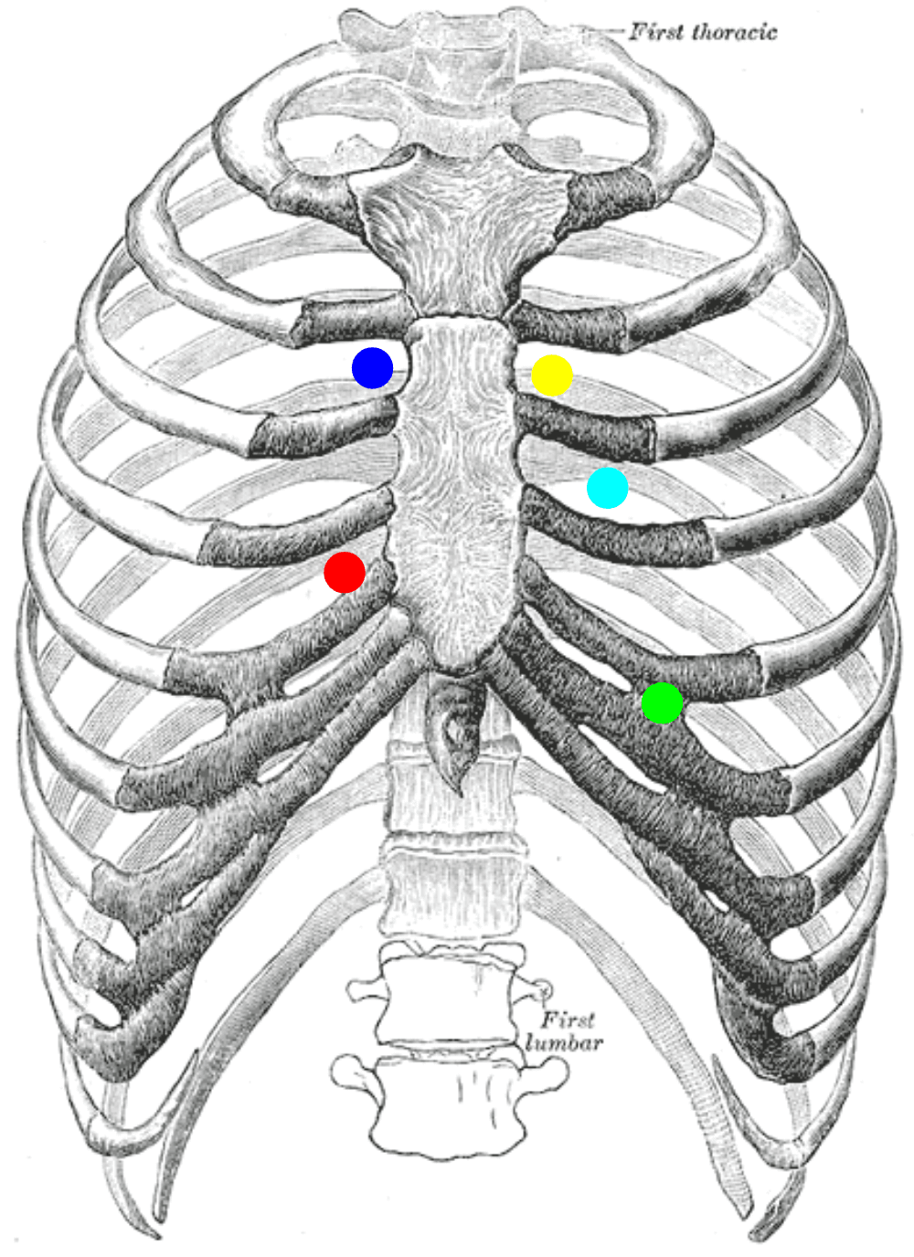
Illustration: heart auscultation areas and common murmurs. Source: Osmosis educational graphic, original post. Use the diagram as a location guide; the written explanations below explain the physiology.
1. First understand what the heart is doing
The heart has four chambers and four valves. The valves act like doors that should open to let blood move forward and close to prevent backward flow.
- Tricuspid valve: right atrium → right ventricle.
- Pulmonary valve: right ventricle → pulmonary artery → lungs.
- Mitral valve: left atrium → left ventricle.
- Aortic valve: left ventricle → aorta → body.
The right heart sends blood to the lungs; the left heart sends blood to the body. The aorta is connected to the left ventricle. It is not “the right side of the heart.” The aortic listening area happens to be on the patient's upper right chest because sound travels through tissues and vessels; it does not mean the aorta is on the right side of the heart.
2. What are S1 and S2?
A normal heartbeat is not one sound. The main sounds are S1 — “lub” and S2 — “dub.”
- S1 (“lub”): closure of the mitral and tricuspid valves. It marks the beginning of ventricular systole.
- S2 (“dub”): closure of the aortic and pulmonary valves. It marks the end of systole and the beginning of ventricular diastole.
Think: S1 — squeeze begins; S2 — squeeze ends. The ventricles contract between S1 and S2; they relax and fill between S2 and the next S1.
A useful pulse trick: feel the carotid pulse gently while listening. Systole occurs around the carotid upstroke, helping you decide whether a sound is systolic or diastolic. Do not compress both carotid arteries.
3. Systole versus diastole: what the words mean
- Systole: ventricular contraction/ejection. On the listening timeline it is between S1 and S2.
- Diastole: ventricular relaxation and filling. It is between S2 and the next S1.
A murmur is described by when it occurs in this cycle. It may be systolic, diastolic, or continuous. It is not enough to say “there is a murmur”; identify its timing and character.
What does “pansystolic” mean?
“Pan” means throughout. A pansystolic (holosystolic) murmur starts with S1 and continues all the way to S2, filling the whole of systole. Classic causes include mitral regurgitation and tricuspid regurgitation; a ventricular septal defect is another classic cause.
A early diastolic murmur begins just after S2, during early ventricular relaxation. Aortic regurgitation classically causes a high-pitched, blowing, decrescendo early-diastolic murmur.
4. Regurgitation versus stenosis
These words describe different mechanical problems.
- Regurgitation (incompetence/insufficiency): the valve does not close properly, so some blood leaks backwards.
- Stenosis: the valve does not open properly, so blood has difficulty moving forwards through a narrowed opening.
The timing depends on when that valve should be closed or open. During ventricular systole, the mitral and tricuspid valves should be closed; if either leaks, blood flows backwards into an atrium and produces a systolic murmur. During diastole, the aortic and pulmonary valves should be closed; if the aortic valve leaks, blood flows back into the left ventricle and produces a diastolic murmur. Mitral stenosis obstructs blood entering the left ventricle during diastole, so it produces a diastolic murmur.
5. The common murmur map
| Lesion | Main murmur timing | Best place to listen | Typical description |
|---|---|---|---|
| Mitral regurgitation | Pansystolic | Apex (mitral area) | Blowing; may radiate to left axilla |
| Tricuspid regurgitation | Pansystolic | Lower left sternal edge | Blowing; often louder with inspiration |
| Aortic stenosis | Ejection systolic | Right 2nd intercostal space, sternal edge | Crescendo–decrescendo; may radiate to carotids |
| Pulmonary stenosis | Ejection systolic | Left 2nd intercostal space, sternal edge | Crescendo–decrescendo |
| Aortic regurgitation | Early diastolic | Left sternal edge, commonly 3rd–4th intercostal space; listen with patient leaning forward | High-pitched blowing, fading away |
| Mitral stenosis | Mid/late diastolic | Apex, often with patient in left lateral position | Low-pitched rumble; may have an opening snap |
This is a learning guide, not a substitute for supervised examination. Murmurs vary with severity, body habitus, heart rate and associated disease; confirm suspected valve disease with echocardiography.
6. Where exactly do I place the stethoscope?
The stethoscope does not sit directly over each valve's anatomical location. These are listening areas, where sound from each valve is transmitted most clearly.
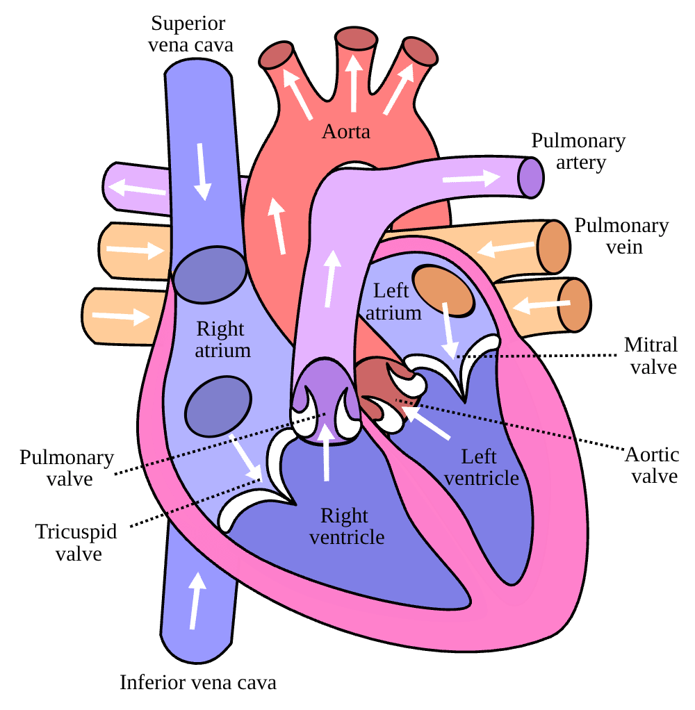
Illustration: auscultation areas. Source: Medbullets heart auscultation diagram.
Use the mnemonic “APE To Man”:
- A — Aortic: 2nd intercostal space, right sternal edge.
- P — Pulmonary: 2nd intercostal space, left sternal edge.
- E — Erb’s point: 3rd intercostal space, left sternal edge; useful for hearing some aortic regurgitation murmurs.
- T — Tricuspid: lower left sternal edge, usually around the 4th–5th intercostal spaces.
- M — Mitral: apex, usually 5th left intercostal space at or near the midclavicular line in an adult.
Intercostal space means the gap between two ribs. Find the sternal angle (Angle of Louis) to identify the 2nd rib, then count down. The exact apex position can vary, especially with cardiomegaly or pregnancy.
Why is aortic regurgitation not necessarily heard on the right?
Because the best listening point is not the same as the physical location of the valve. Aortic regurgitation is often best heard along the left sternal edge (including Erb’s point), especially with the patient sitting forward at end-expiration. The aortic area on the right upper sternal edge is a standard area for aortic stenosis and for systematically listening to the aortic valve, but it is not the only place to listen for aortic valve disease.
7. How to perform a basic cardiovascular examination
Explain the examination, obtain consent, wash hands, position the patient at about 45 degrees, expose the chest appropriately while maintaining dignity, and ensure good lighting.
A. General inspection and hands
Look for distress, pallor, central/peripheral cyanosis, scars, oxygen, and signs of infective endocarditis or chronic disease. Inspect the fingers for clubbing, splinter haemorrhages, and peripheral perfusion. Osler nodes are tender nodules on finger/toe pads; Janeway lesions are classically painless lesions on palms/soles. These signs are not always present and are not diagnostic alone.
B. Pulse and blood pressure
Assess pulse rate, rhythm, volume and character; compare both radial pulses when appropriate. Measure blood pressure correctly. A collapsing/bounding pulse and wide pulse pressure may support significant aortic regurgitation but are not diagnostic by themselves.
C. Face and neck
Look for pallor, cyanosis and xanthelasma where relevant. Assess the jugular venous pressure (JVP) with the patient at 45 degrees, distinguishing the venous waveform from the carotid pulse. Raised JVP can suggest elevated right-sided filling pressure, including right heart failure.
D. Precordium
Inspect for scars, visible pulsations, deformity or a pacemaker. Palpate for the apex beat, heaves and thrills. A thrill is a palpable vibration caused by turbulent flow. Locate the apex beat and assess whether it is displaced; a displaced apex may indicate cardiac enlargement but must be interpreted in context.
E. Auscultation
Listen systematically at all five areas. At each point ask:
- Can I identify S1 and S2?
- Is the rhythm regular?
- Are there extra sounds, such as S3, S4 or a click?
- Is there a murmur? If so, what is its timing, pitch/character, loudness, location and radiation?
Use the diaphragm for most higher-pitched sounds. Use the bell lightly for low-pitched sounds such as the rumble of mitral stenosis. If you hear a murmur, listen to areas where it may radiate: the carotids for aortic stenosis and the left axilla for mitral regurgitation. Listen in different positions when appropriate.
F. Look for heart failure
Assess respiratory rate, oxygen saturation, lung bases for crackles, JVP, ankle/sacral oedema, and the abdomen for hepatomegaly or ascites. Do not diagnose heart failure from one sign alone.
8. The link to acute rheumatic fever
Acute rheumatic fever can follow group A streptococcal infection and cause inflammation of the heart (carditis). Inflammation can affect the endocardium and valves. Chronic rheumatic heart disease particularly affects the mitral valve, and may also affect the aortic valve.
- Mitral regurgitation: the mitral valve leaks during systole. A pansystolic murmur is classically heard at the apex and may radiate to the left axilla.
- Aortic regurgitation: the aortic valve leaks during diastole. A high-pitched early diastolic murmur is classically heard along the left sternal edge.
Severe valve dysfunction can increase pressure in the lungs and/or cause reduced forward cardiac output. Look for tachypnoea, breathlessness, crackles, raised JVP, peripheral oedema and hepatomegaly. In a child or young adult with suspected acute rheumatic fever and carditis, urgent clinical assessment and echocardiography are important. A murmur may be subtle or absent despite carditis, so a normal-sounding examination does not rule it out.
9. How to answer “What did you hear?”
Do not jump straight to naming a valve lesion. Describe what you actually heard first.
If normal, say:
“On auscultation, S1 and S2 are audible, the rhythm is regular, and I hear no added sounds or murmurs.”
Only say “no murmurs” if you have listened systematically and are confident. If you cannot identify the sounds, say so honestly and ask to repeat under supervision.
If you suspect a murmur, use this order:
- Timing: systolic, diastolic or continuous.
- Duration/shape: pansystolic, early diastolic, ejection systolic, etc.
- Character/pitch: blowing, harsh, rumbling.
- Maximum intensity: apex, right upper sternal edge, left sternal edge, etc.
- Radiation: axilla, carotids or elsewhere.
- Grade and associated signs, if you can assess them reliably.
Example: “There is a high-pitched, blowing, early-diastolic murmur best heard at the left sternal edge with the patient sitting forward. This is suggestive of aortic regurgitation; I would correlate with the rest of the examination and echocardiography.”
10. Rapid recall
- S1: mitral + tricuspid closure; start of systole.
- S2: aortic + pulmonary closure; end of systole/start of diastole.
- MR and TR: classically pansystolic.
- AR: classically early diastolic.
- MS: classically mid-diastolic rumble.
- Aortic area: right 2nd intercostal space.
- Pulmonary area: left 2nd intercostal space.
- Mitral area: apex, left 5th intercostal space near midclavicular line.
- Aortic regurgitation is often best heard at the left sternal edge, not only at the right-sided aortic area.
Further reading
- American Heart Association: Heart Valve Auscultation Guide
- NCBI Bookshelf: Cardiovascular examination
- Merck Manual: Cardiac auscultation
Next: Respiratory Examination and Chest Findings.
11. Extra bedside details that examiners commonly expect
Pulse: what to describe
Count the rate (typically 60–100 beats/min in a resting adult), assess rhythm, volume and character, and compare both radial pulses when indicated. Check for radio-radial delay and, when clinically indicated, radio-femoral delay. A collapsing pulse can occur in significant aortic regurgitation; a slow-rising pulse can occur in aortic stenosis. These are clues, not diagnoses. Measure blood pressure and consider both arms when appropriate.
JVP: the simple principle
The jugular venous pressure estimates right atrial pressure. Examine with the patient reclined around 45 degrees, head turned slightly away, using tangential light to identify the internal jugular venous pulsation. It is normally assessed vertically above the sternal angle; use your school's accepted measurement method and reference point. The venous waveform is usually not palpable and changes with posture and respiration, unlike the carotid pulse. A raised JVP may occur in right heart failure, fluid overload, tamponade or constrictive pericarditis; interpret with the rest of the examination.
Apex beat, heaves and thrills
The apex beat is the most lateral/inferior palpable impulse from the left ventricle. Locate it with the patient at 45 degrees and, if not felt, consider the left lateral position. A displaced or diffuse apex can suggest enlargement. A heave is a sustained lifting impulse, which may reflect ventricular hypertrophy. A thrill is a palpable murmur vibration. Do not confuse a thrill (palpable) with a murmur (heard).
Added heart sounds
- S3: occurs just after S2 in early diastole. It can be physiological in children, young adults and pregnancy, but in an older adult may suggest volume overload or heart failure.
- S4: occurs just before S1, during atrial contraction, and may indicate a stiff ventricle; it is absent in atrial fibrillation.
- Opening snap: may occur after S2 in mitral stenosis.
- Ejection click: may occur with some abnormal semilunar valves.
Listen with the diaphragm and bell as appropriate, and describe what you hear rather than forcing a diagnosis.
A complete presentation
“On cardiovascular examination, the patient is [comfortable/distressed]. The pulse is ___ beats/min, [regular/irregular], with ___ volume; blood pressure is ___. JVP is ___. The apex beat is ___. Heart sounds S1 and S2 are ___. There is [no murmur / a murmur described by timing, character, site and radiation]. The lung bases show ___, and there is [no/present] peripheral oedema or hepatomegaly.” Only include observations you actually assessed.
12. Starting the cardiovascular OSCE and general inspection
Use the same professional opening as other examinations: clean hands; introduce yourself; confirm the patient's identity; explain the examination and obtain consent; ask about pain; position the patient at approximately 45 degrees; expose the chest appropriately while maintaining dignity. If the patient is acutely unwell, assess ABCs and call for help rather than continuing a routine exam.
General inspection, hands, nails, face and mouth
- General appearance: breathlessness, pallor, cyanosis, sweating, cachexia, oxygen, mobility aids and signs of distress.
- Hands/nails: peripheral temperature and perfusion, capillary refill, clubbing, splinter haemorrhages, nicotine staining, palmar erythema and signs of anaemia. Osler nodes are tender; Janeway lesions are classically painless. These infective-endocarditis signs may be absent and are not diagnostic alone.
- Pulse: rate, rhythm, volume and character; compare radial pulses and assess radio-radial/radio-femoral delay if indicated.
- Face/mouth: conjunctival pallor, central cyanosis (look at the tongue/oral mucosa), xanthelasma and dental/oral health where relevant.
- Neck: JVP at about 45 degrees; assess carotid pulse only when appropriate and one side at a time. Never palpate both carotids simultaneously.
- Precordium: inspect for scars, deformity, devices and visible pulsations before palpation and auscultation.
A reliable sequence
General inspection → hands and pulse/BP → face → JVP and neck → precordium (inspect, palpate apex/heaves/thrills, auscultate all areas) → lungs for crackles → ankles/sacrum for oedema → abdomen for hepatomegaly/ascites when indicated. Local OSCE sequences can vary; state what you did and do not invent findings.
Vascular bruits: where they fit
A bruit is a blowing/whooshing sound from turbulent blood flow. It differs from a heart murmur mainly by the site and context; both are sounds of turbulent flow.
- Carotid bruit: listen over each carotid artery separately when indicated, with the patient comfortable. Avoid pressing hard with the stethoscope and do not auscultate both sides simultaneously.
- Abdominal aortic bruit: listen over the midline upper abdomen.
- Renal artery bruits: listen in the upper abdomen, slightly to the right and left of the midline, above/near the umbilicus along the renal artery course. A bruit may raise suspicion of renal artery stenosis in a patient with resistant hypertension, but absence does not exclude it and presence does not prove it.
- Iliac bruits: listen lower in the abdomen toward the right and left iliac vessels if peripheral arterial disease is suspected. Anatomically, the common iliac arteries divide into internal and external iliac arteries; do not call these renal arteries.
- Femoral bruits: listen in the groin over each femoral artery if indicated.
A bruit is not routinely required in every basic cardiac station. Follow the examiner's instruction and the clinical indication; if the task is a full cardiovascular/peripheral vascular examination, know where it belongs.