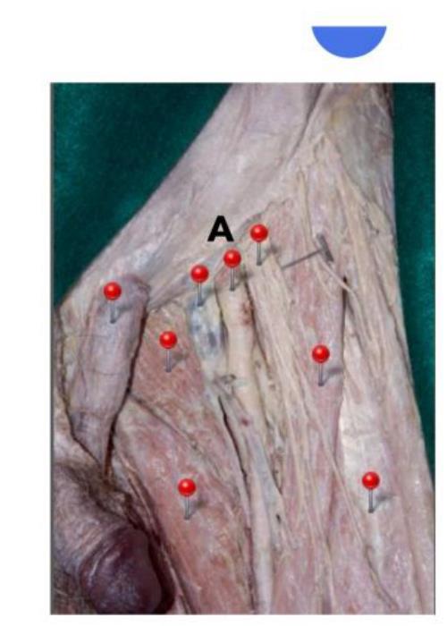
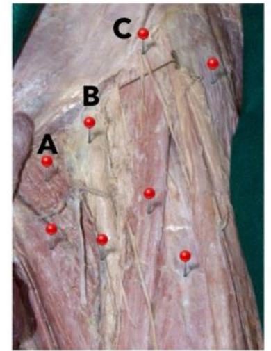
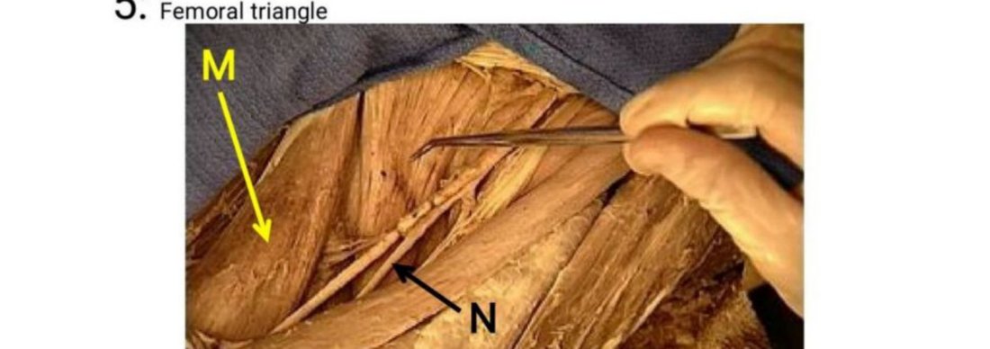
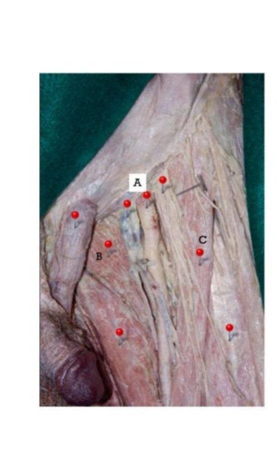
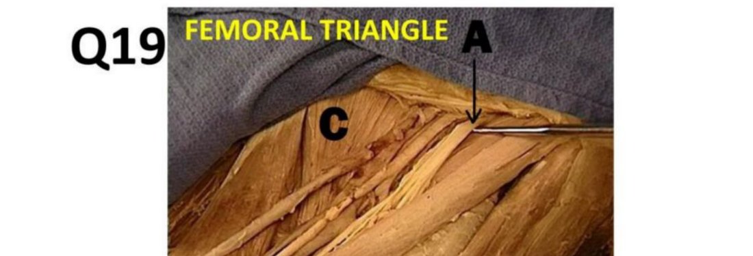
Femoral Triangle
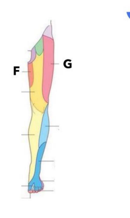
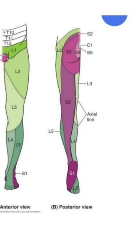
Q63: State the dermatomes of the femoral triangle and anterior knee
What our notes say
- Q63: State the dermatomes of the femoral triangle and anterior knee (Aponeurosis — Anatomy Question Bank)
- Q11 — LOWER LIMB & DEVELOPMENTa–b) Femoral Triangle — see Q4b above. (Master First-Year Anatomy: Must-Know Essays & Key Concepts)
- Femoral vein: ascends femoral triangle; receives deep femoral vein and great saphenous vein; becomes external iliac vein at inguinal ligament (Master First-Year Anatomy: Must-Know Essays & Key Concepts)
Study material on Femoral Triangle
Images
Practice questions on Femoral Triangle
Regarding the femoral triangle
- The lateral border of the adductor longus makes up its medial boundary
- It is bisected by the femoral nerve
- The lateral border of sartorius muscle makes up its lateral boundary
- The long saphenous vein joins the femoral vein within the triangle
- Rectus femoris makes up part of the floor
Answer: A. The great (long) saphenous vein pierces the cribriform fascia and joins the femoral vein within the femoral triangle at the saphenous opening. ---
Which artery is a direct branch of the femoral artery, typically arising proximally in the femoral triangle?
- Obturator artery
- Perforating artery
- Profunda femoris artery
- Inferior epigastric artery
Answer: C. The profunda femoris (deep femoral) artery is the largest branch of the femoral artery, typically arising about 3.5 cm below the inguinal ligament. Perforating arteries are branches of the profunda femoris artery. The obturator and inferior epigastric arteries are branches of the internal and external iliac arteries, respectively.