Femur
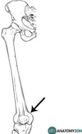
• Is relatively easily palpated against the underlying femur
What our notes say
- • Is relatively easily palpated against the underlying femur (Cardiovascular system examination revised 2012 — Past Paper Questions & Answers)
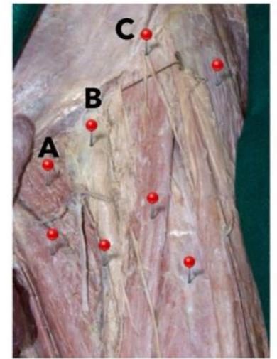
- Surface landmarks: patella (kneecap), patellar tendon, medial joint line, lateral joint line, quadriceps muscle, hamstring muscle group, tibia, anterior tibial tuberosity (insertion of the patellar tendon), and femur. (muscuskeletal exam — Past Paper Questions & Answers)
- The medial and lateral menisci sit on top of the tibia, providing a cushioned articulating surface between the femur and tibia. (muscuskeletal exam — Past Paper Questions & Answers)
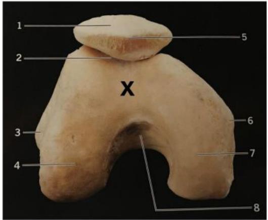
- A 21 year old male had a resection of the distal Femur of the right (pathology essays — Past Paper Questions & Answers)
- Clinical relevance: risk of avascular necrosis after a fracture of the neck of femur (Aponeurosis — Anatomy Question Bank)
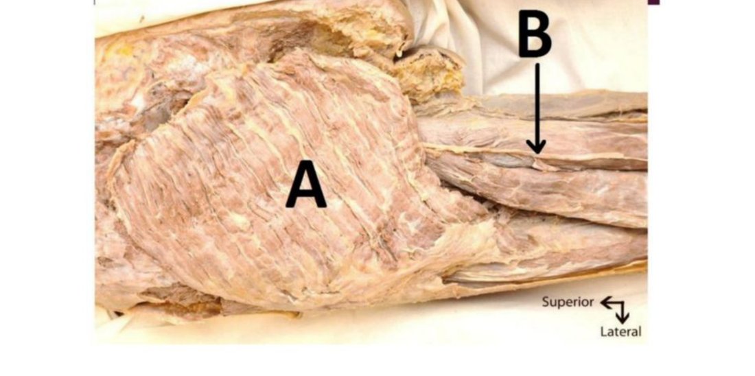
- Prevents forward dislocation of the femur while crouching (Aponeurosis — Anatomy Question Bank)
Study material on Femur
- muscuskeletal exam
- Bone & Cartilage Tumors: Osteoma to Osteosarcoma Pathology
- Aponeurosis — Anatomy Question Bank
- Master Bone & Soft Tissue Pathology: CAT 2 MCQs & Answers
- Bone & Soft Tissue Pathology: Cases, Tumors, & Answers
- Bone &Soft Tissue yr3 2025 — Past Paper Questions & Answers
Images
Practice questions on Femur
A 17-year-old boy presents with progressive pain and swelling around the distal femur. Radiography demonstrates a destructive mixed lytic and sclerotic lesion with a Codman triangle. What is the most likely diagnosis?
- Osteochondroma
- Osteoid osteoma
- Osteosarcoma
- Chondroma
- Giant cell tumour ✅ Answer: C
Answer: A. Codman triangle, mixed lytic/sclerotic lesion, and presentation in a young patient are classic features of osteosarcoma.
A 19-year-old man has a painless bony outgrowth arising from the metaphysis of the distal femur. Radiography shows continuity of the medullary cavity with the host bone. What is the most likely diagnosis?
- Osteochondroma
- Enchondroma
- Osteoblastoma
- Osteosarcoma
- Fibrous dysplasia ✅ Answer: D
Answer: A. A painless bony outgrowth from the metaphysis with continuity of the medullary cavity and cortex with the host bone is the classic description of an osteochondroma (A). Osteosarcoma (D) is a malignant tumor and would not typically present with these radiographic features.
The 'screw-home' mechanism of the knee joint involves passive medial rotation of the femur on the tibia during terminal extension. This mechanism causes tightening of which primary ligament, contributing to knee stability?
- Fibular (lateral) collateral ligament
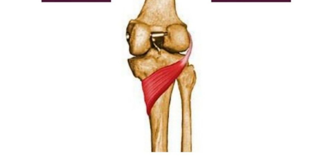
- Tibial (medial) collateral ligament
- Posterior cruciate ligament
- Patellar ligament
Answer: B. The screw-home mechanism tightens both the collateral and cruciate ligaments, but the tibial (medial) collateral ligament is particularly important in resisting external rotation and providing stability in full extension, along with the anterior cruciate ligament. The question asks for a primary ligament that becomes tight due to the mechanism, and the MCL is a strong contributor to full extension stability.