Hyperplasia
Next, an endometrial biopsy is performed, and there is microscopic evidence for endometrial hyperplasia.
What our notes say
- Next, an endometrial biopsy is performed, and there is microscopic evidence for endometrial hyperplasia. (Female system — Past Paper Questions & Answers)
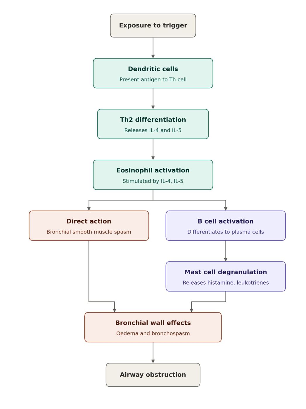
- Pathology: mucous gland hypertrophy and goblet cell hyperplasia (increased Reid index above 0.5), mucus hypersecretion, chronic inflammation, ciliary dysfunction, bronchial wall thickening and airway narrowing. (Bronchitis (Acute and Chronic))
- The presence of endometrial glands and stroma within the myometrium, with surrounding smooth muscle hyperplasia. (Year 4 Gynaecology CAT 2 (MBOG 4212): 8 October 2021)
- No increased risk (non-proliferative): cysts, simple fibroadenoma, duct ectasia, mild hyperplasia. (General Surgery CAT 2 (MBSG 4600): 4 October 2021)
- Slightly increased risk (about 1.5 to 2 times; proliferative without atypia): moderate or florid hyperplasia, sclerosing adenosis, intraductal papilloma and complex fibroadenoma, radial scar. (General Surgery CAT 2 (MBSG 4600): 4 October 2021)
- SP-B, SP-C, ABCA3 and NKX2-1 defects and neuroendocrine cell hyperplasia of infancy cause childhood ILD. (Paediatrics end of 2nd semester exam (MBPE 4300): 5 October 2021, 50 MCQs)
Study material on Hyperplasia
- Exam Prep: Skin Infections, Psoriasis & Pemphigus
- ENDOCRINE PATHOLOGY
- Female system
- Female Reproductive Pathology: HPV, Cancer, PCOS & Pregnancy
- General Pathology MCQs: Fixatives, FNA, Endocrine & ARDS
- Disorders of the Vagina and Breast
- Chemical pathology and Endocrine pathology
- Benign Breast Epithelial Lesions: Types, Risk & Pathology
- Dr. Irungu Notes
- White Cell Disorders — Must-Know Notes
- Hematopathology Mcqs 2
- Pituitary + Thyroid & Parathyroid
Images
Practice questions on Hyperplasia
A 72-year-old man has progressive fatigue over 2 years. Bone marrow biopsy shows hypercellularity with dysplastic megakaryocytes, erythroid hyperplasia, and 6% blasts. Cytogenetics shows deletion of chromosome 5q. Most likely prognosis?
- Very poor — likely transforms to AML within weeks
- Good — 5q deletion is associated with favorable prognosis in MDS
- Intermediate — requires immediate bone marrow transplant
- Poor — 5q deletion always indicates secondary AML
Answer: B. Isolated 5q deletion in MDS (5q- syndrome) is associated with a favorable prognosis, macrocytic anemia, low blast count, and good response to lenalidomide. It is one of the best prognostic cytogenetic findings in MDS. ---
A 40-year-old man has a peripheral blood eosinophil count of 4500/microliter persisting for 6 months. He has no parasitic infection, no allergic disease, and no drug exposure. Bone marrow shows eosinophilic hyperplasia. FISH reveals FIP1L1-PDGFRA fusion gene. Most likely diagnosis?
- Reactive eosinophilia from parasitic infection
- Hypereosinophilic syndrome
- Eosinophilic leukemia with PDGFRA rearrangement
- Allergic bronchopulmonary aspergillosis
Answer: C. Persistent marked eosinophilia + FIP1L1-PDGFRA fusion gene = eosinophilic leukemia. This is a clonal myeloid neoplasm driven by a tyrosine kinase fusion. Unlike reactive eosinophilia, it has a primary clonal driver and responds to imatinib. ---
A 60-year-old woman has had headaches and visual disturbances for 3 months. Platelet count is 1,200,000/microliter. WBC and hemoglobin are normal. Bone marrow shows megakaryocytic hyperplasia with large clustered megakaryocytes. JAK2 mutation is present. Most likely diagnosis?
- Reactive thrombocytosis from iron deficiency
- Myelofibrosis
- Chronic myelogenous leukemia
- Essential thrombocythemia
Answer: D. Markedly elevated platelets + normal WBC and Hgb + megakaryocytic hyperplasia + JAK2 mutation + thrombotic symptoms = essential thrombocythemia. Reactive thrombocytosis does not cause symptoms and JAK2 is negative. ---
A 35-year-old woman has a 3 cm firm non-tender left axillary lymph node. Biopsy shows follicular hyperplasia with prominent germinal centers, polyclonal B and T cells, plasma cells, and macrophages. No Reed-Sternberg cells. No monoclonal population on flow cytometry. Most likely diagnosis?
- Follicular lymphoma
- Reactive follicular hyperplasia
- Hodgkin lymphoma
- Marginal zone lymphoma
Answer: B. Prominent germinal centers + polyclonal mixed population + no monoclonal cells = reactive follicular hyperplasia. Unlike follicular lymphoma (monoclonal, crowded nodules, BCL2+), reactive hyperplasia has a mixed polyclonal population with preserved architecture. ---
Water-clear cell hyperplasia refers to
- Oxyphil cell adenoma with clear cytoplasm
- Secondary hyperplasia cells in CRF
- Parathyroid carcinoma with vacuolated cells
- Rare form of primary hyperplasia where cells have abundant clear glycogen-rich cytoplasm
Answer: D. Water-clear cell (wasserhelle cell) hyperplasia is a rare form of primary hyperplasia. All four glands are massively enlarged (can reach >10g total). Cells have large, empty-looking cytoplasm due to glycogen and vacuoles. Associated with marked hypercalcemia. ---
In primary parathyroid hyperplasia, the combined weight of all four glands typically
- Is less than 1g total
- Varies unpredictably between glands
- Matches normal gland weight (140–160mg total)
- Exceeds 1g total (normal ~35–40mg each)
Answer: D. Normal total parathyroid weight = ~140–160mg (4 glands × ~35–40mg each). In primary hyperplasia, total gland weight exceeds 1g. In secondary hyperplasia from CRF, combined weight can reach several grams. All four glands are involved though asymmetrically. ---