Pectoralis Major
The chest tube is placed (on the correct side) in the mid- or anterior- axillary line, behind pectoralis major (to avoidhaving to dissect through this thick muscle).
What our notes say
- The chest tube is placed (on the correct side) in the mid- or anterior- axillary line, behind pectoralis major (to avoidhaving to dissect through this thick muscle). (Examination of Respiratory system — Past Paper Questions & Answers)
- Safe triangle: pectoralis major, latissimus dorsi, 5th intercostal space. (Pneumothorax: Classification, Clinical Features and Emergency Management)
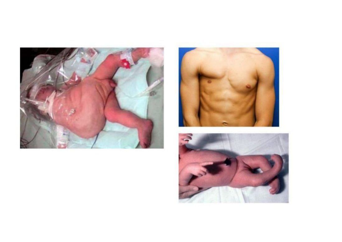
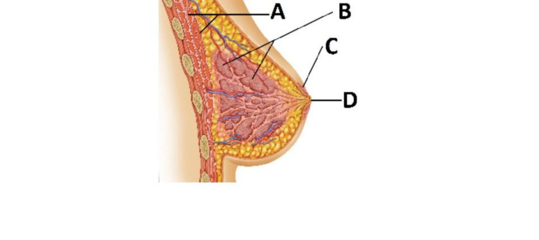
- Poland syndrome — absent pectoralis major with breast and nipple hypoplasia (Aponeurosis — Embryology Question Bank)
- Associated anomaly: Poland's syndrome (congenital absence/underdevelopment of the pectoralis major, often with hand anomalies) (Aponeurosis — Histology Question Bank)
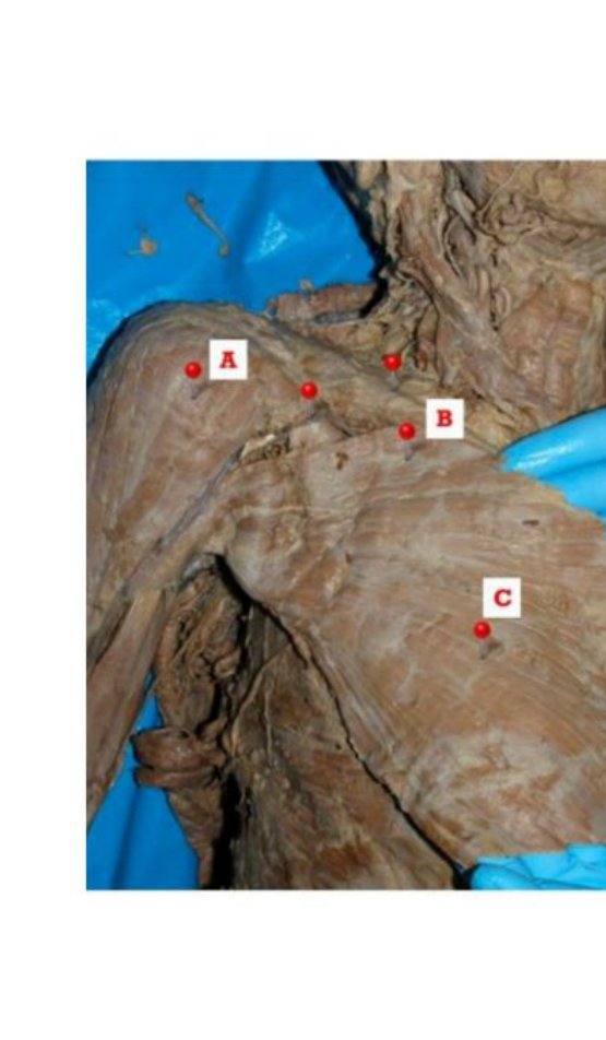
- Two actions of C (pectoralis major) on the arm: adduction; medial rotation (Aponeurosis — Anatomy Question Bank)
Study material on Pectoralis Major
Images