Intervertebral Disc
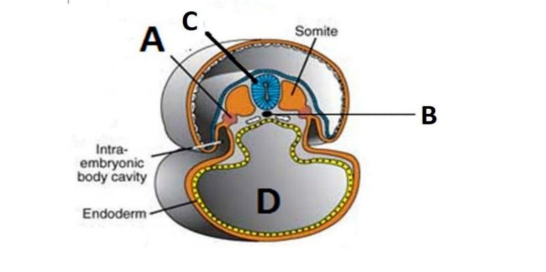
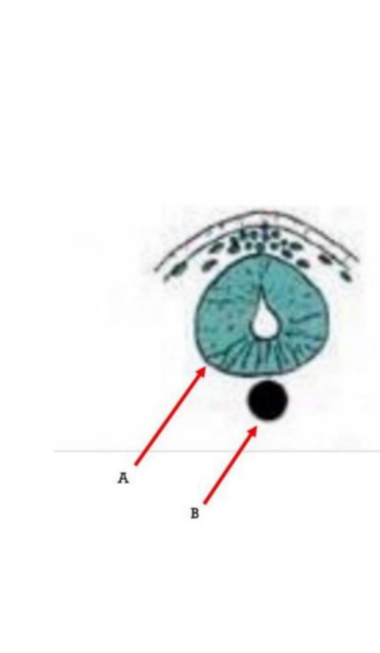
Derivative: nucleus pulposus of the intervertebral disc.
What our notes say
- Derivative: nucleus pulposus of the intervertebral disc. (Aponeurosis — Embryology Question Bank)
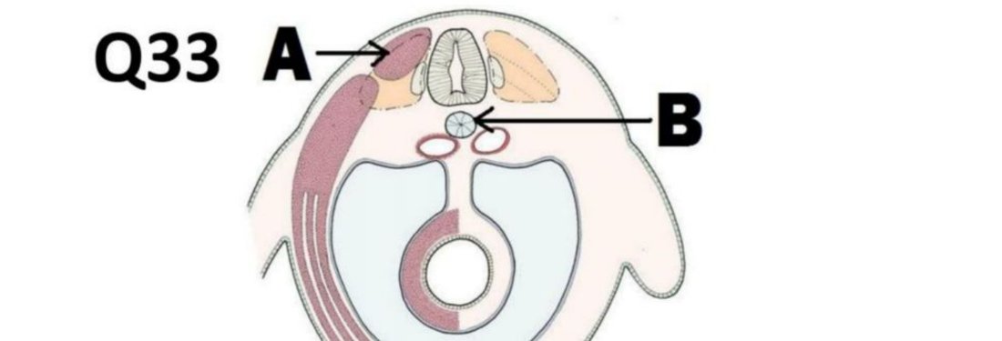
- Two fates of B (notochord): mostly regresses; the remnant persists as the nucleus pulposus of the intervertebral disc (Aponeurosis — Embryology Question Bank)
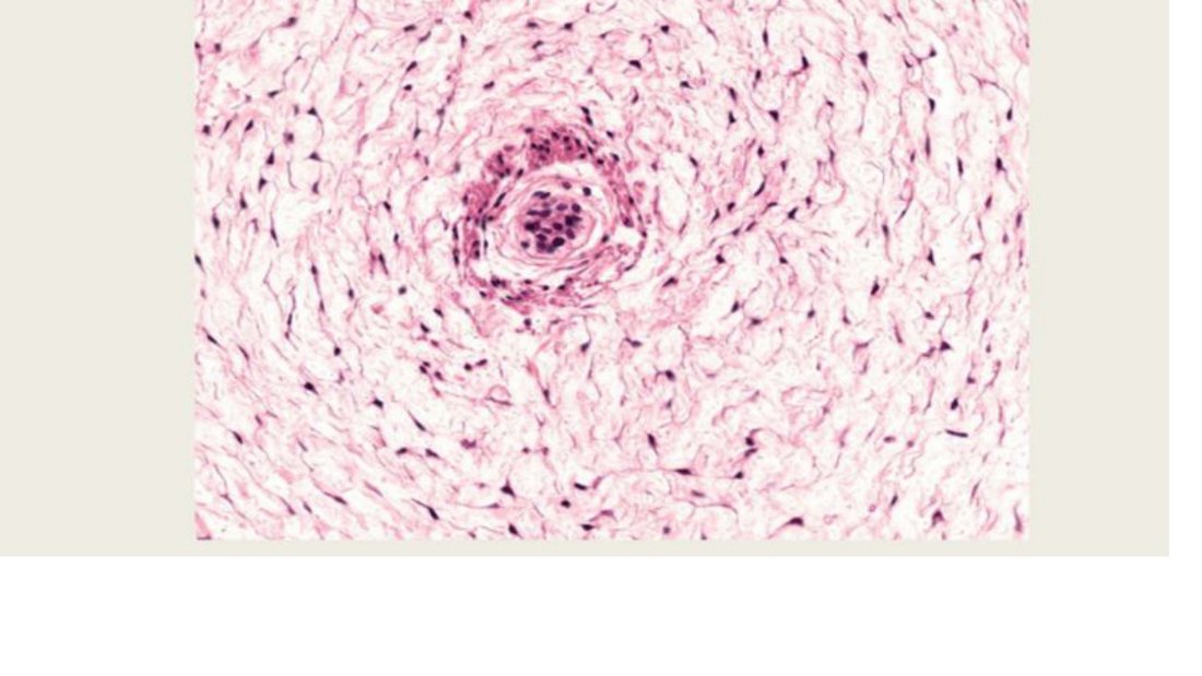
- Found in articular discs, symphyses, intervertebral discs, and tendon-to-bone attachments; resists compression and tensile stress (Aponeurosis — Histology Question Bank)
- Three sites where found: knee menisci; intervertebral discs; pubic symphysis (Aponeurosis — Histology Question Bank)
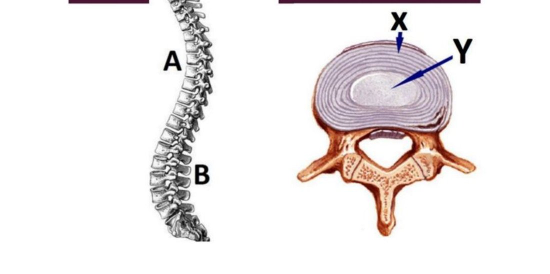
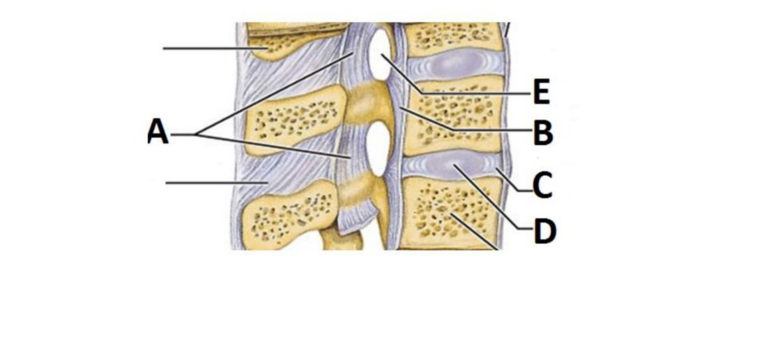
- Q92: Outline four actions of the intervertebral disc, A (4 marks) (Aponeurosis — Anatomy Question Bank)
- 40% of cases involve the spine (Pott disease) — infection crosses intervertebral discs, destroys vertebrae + soft tissue → compression fractures → scoliosis, kyphosis, neurologic deficits (Bone & Joint Pathology: Basic Structure, Cells, and Fractures)
Study material on Intervertebral Disc
Images
Practice questions on Intervertebral Disc
The internal iliac artery is crossed by which of the following structures at the level of the intervertebral disc between L5 and S1? ---
- Puborectalis muscle
- Ureter
- Obturator nerve
- Umbilical artery
Answer: B.