Skull
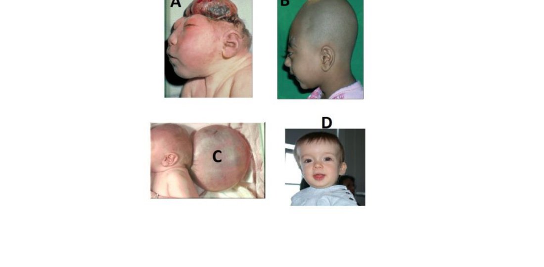
With the lemon sign, the frontal bones are flattened (almost concave) so that the anterior skull is not as rounded.
What our notes say
- With the lemon sign, the frontal bones are flattened (almost concave) so that the anterior skull is not as rounded. (CNS PATHOLOGY — Past Paper Questions & Answers)
- However, an hour later in the emergency room, he is comatose, and skull radiograph reveals a linear skull fracture of the temporal-parietal region on the left. (CNS PATHOLOGY — Past Paper Questions & Answers)
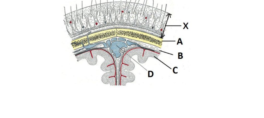
- Epidural haematoma is due to rapid accumulation of blood between the dura matter and skull (Behavioural Sciences MCQs — December 2013 Paper I (Past Paper))
- The brain and spinal cord are enclosed in a rigid, non-expansile compartment (skull + dural reflections + bony spinal canal). (Cerebral Edema, Hydrocephalus, Raised ICP & Brain Herniation Pathology)
- Musonge, 5 years, has recurrent yellowing of the eyes, three blood transfusions and skull bossing. (Paediatrics end of fourth year main exam: 1 July 2019, Paper One, 99 of 100 MCQs)
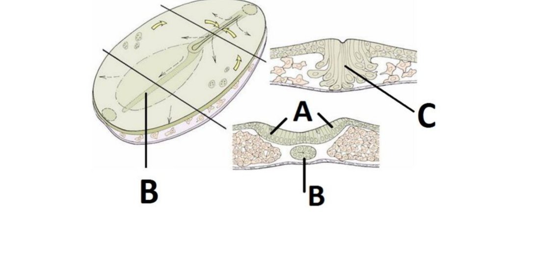
- Typically found along the midline, from skull base to sacrum (Aponeurosis — Embryology Question Bank)
Study material on Skull
- CNS Trauma Neuropathology: Fractures, Concussion & CTE
- CNS PATHOLOGY
- Aponeurosis — Embryology Question Bank
Images
Practice questions on Skull
A 72-year-old man presents with back pain and confusion. X-ray shows punched-out lytic skull lesions. Serum electrophoresis shows an IgG M protein spike. Urine shows Bence Jones proteins. Bone marrow has >30% plasma cells. Creatinine is markedly elevated. Which mechanism BEST explains his renal dysfunction?
- IgG deposition causing membranous nephropathy
- Hypocalcaemia causing renal vasoconstriction
- Obstructive casts of Bence Jones proteins in distal tubules causing tubular necrosis
- Lymphomatous infiltration of the renal parenchyma
Answer: C. This is multiple myeloma. Renal dysfunction results primarily from obstructive proteinaceous casts — composed mostly of Bence Jones proteins (free light chains) along with Tamm-Horsfall protein and albumin — forming in the distal convoluted tubules and collecting ducts. Adjacent epithelial cells become necrotic from the toxic effects of Bence Jones proteins. It is hypercalcaemia (not hypocalcaemia) that also contribu
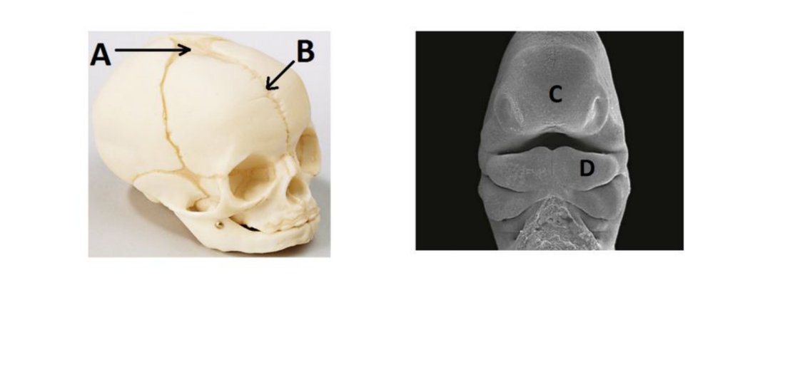
Which of the following is MOST CORRECT regarding the formation of the skull?
- The neurocranium has a membranous part which undergoes endochondral ossification
- The neurocranium has a membranous part which undergoes both endochondral and intramembranous ossification
- The base of the skull arises from 2 sources both of which undergo endochondral ossification
- The base of the skull and the membranous part both undergo intramembranous ossification
- None of the above is correct
Answer: C. ** The skull base develops from both neural crest cells and paraxial mesoderm, and both undergo endochondral ossification. The calvarium undergoes intramembranous ossification. --- ---
A port wine stain in the distribution of the trigeminal nerve is found in a child with seizures, intellectual disability, and skull "tram-track" calcifications. What underlying mutation causes this syndrome?
- Germline VHL mutation causing haemangioblastomas and AV malformations
- TSC1/TSC2 mutation causing tuberous sclerosis complex with cortical tubers
- NF1 mutation causing neurofibromas and café-au-lait macules
- Somatic GNAQ missense mutation in the Gα subunit causing constitutive signalling
Answer: D. Sturge-Weber syndrome (port wine stain + leptomeningeal angiomas + seizures + intellectual disability) is caused by a somatic GNAQ missense mutation occurring early in development — not inherited. ---