Embolism
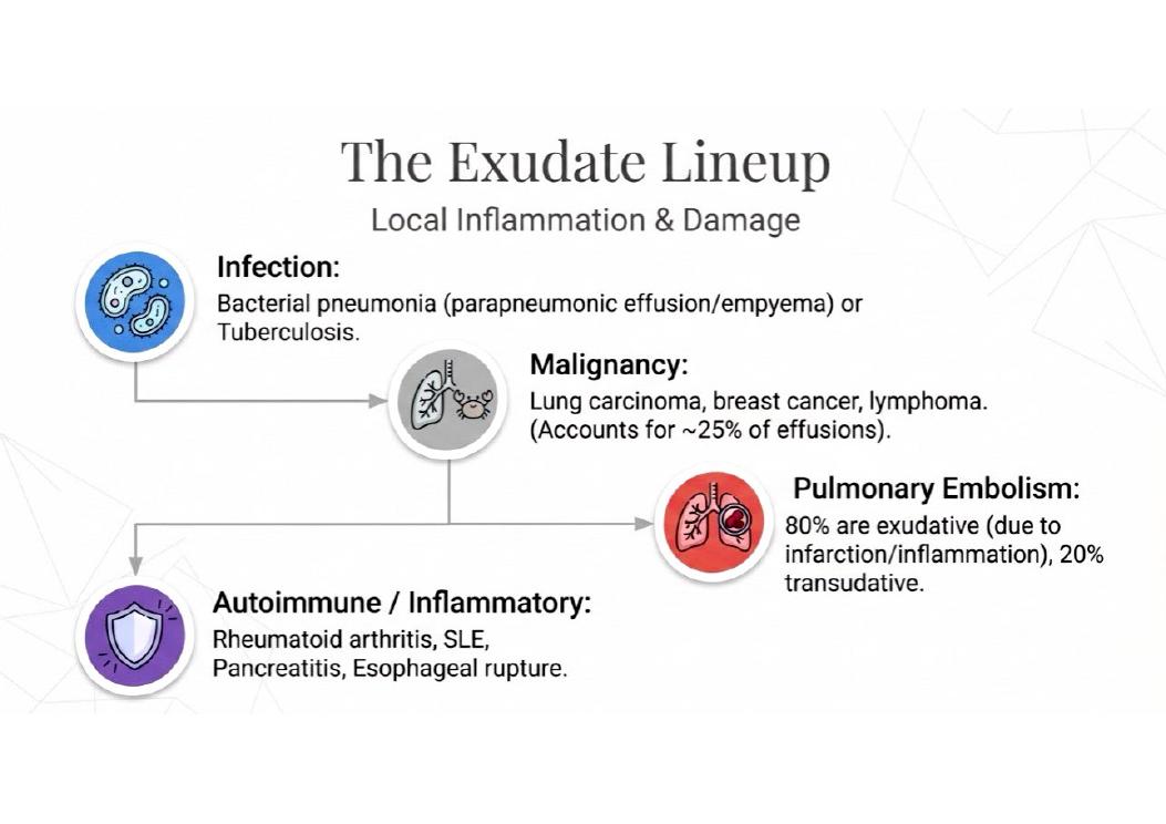
Effusions in pulmonary embolism may be either type, and are often blood-stained.
Embolism (most common)
- Most common territory affected: MCA — equal in both hemispheres
- Emboli lodge at branch points or areas of preexisting stenosis
- Fat embolism → "shower embolization" → diffuse dysfunction + hemorrhagic white matter lesions
Read this in Cerebrovascular Disease: Stroke, Ischemia & Hemorrhage Explained
What our notes say
- Effusions in pulmonary embolism may be either type, and are often blood-stained. (Internal Medicine Respiratory CAT 1: 30 MCQs and 5 short answer questions)
- The target INR is 2 to 3 (aim 2.5) for at least 3 months in a provoked pulmonary embolism. (Internal Medicine (MBIM 4100) main exam, Paper One: 3 July 2019)
- The Wells score is for pulmonary embolism and deep vein thrombosis. (Respiratory Medicine Question Bank: 15 Clinical MCQs)
- BNP is raised in kidney failure, atrial fibrillation, older age and pulmonary embolism, and lower in obesity. (Heart failure: which tests and why)
- Deep Vein Thrombosis and Venous Thromboembolism (with Pulmonary Embolism) — Year 4 Internal Medicine Notes (DVT and Venous Thromboembolism (with Pulmonary Embolism))
- Pulmonary embolism (PE): a clot (usually from a leg DVT) lodges in the pulmonary arteries. (DVT and Venous Thromboembolism (with Pulmonary Embolism))
Study material on Embolism
- DVT and Venous Thromboembolism (with Pulmonary Embolism)
- Cat. Pathology: Acute Inflammation, Mediators & Pulmonary Embolism
- Respiratory Pathology 1b
- Cardiovascular system examination revised 2012
- Examination of Respiratory system
- Junior Clerkship in Internal Medicine (MBIM4100) main exam, Paper One: 31 January 2024
- VASCULAR PATHOLOGY — Must-Know Notes
- Lung Pathology — Must-Know
- Internal Medicine Respiratory CAT 1: 30 MCQs and 5 short answer questions
- Respiratory Medicine Question Bank: 15 Clinical MCQs
- Respiratory System Pathology 1A — Practice Questions
- Respiratory Pathology 2
- Respiratory Pathology
Images
Practice questions on Embolism
A pulmonary infarct after embolism has which morphologic shape?
- Round, centrally located, hemorrhagic nodule
- Wedge-shaped, base at pleura, apex toward hilum
- Diffuse bilateral consolidation of lower lobes
- Irregular cavitary lesion with necrotic center
Answer: B. Pulmonary infarcts are characteristically wedge-shaped with the base at the pleural surface and apex pointing toward the hilum. About 3/4 affect the lower lobes and more than half are multiple. ---
In pulmonary embolism, infarction occurs in only about 10% of cases because
- Most emboli are too small to cause significant obstruction
- The lung has dual blood supply from pulmonary and bronchial arteries
- Fibrinolysis rapidly resolves most emboli before infarction
- Collateral circulation from adjacent lung segments prevents necrosis
Answer: B. The lung is protected from infarction by its dual blood supply: pulmonary arteries, bronchial arteries, and direct oxygenation from alveolar air. Infarction only occurs when this is compromised. ---
Fat embolism syndrome typically develops within what time frame after long bone fracture?
- Immediately within 1–2 hours of injury
- Within 24–72 hours after injury
- Between 1 and 2 weeks after injury
- More than 4 weeks after the initial trauma
Answer: B. Fat embolism syndrome (petechiae, neurologic symptoms, respiratory failure) typically develops 24–72 hours after massive trauma or long bone fracture. The mechanism involves mechanical obstruction plus toxic fatty acid release causing endothelial damage. ---
Amniotic fluid embolism is a catastrophic obstetric emergency primarily because it causes
- Massive pulmonary infarction from large vessel occlusion
- Sudden dyspnea, hypotension, hypoxia, and DIC
- Right heart failure from chronic pulmonary hypertension
- Granulomatous pneumonitis from fetal squamous cells
Answer: B. Amniotic fluid embolism presents with sudden dyspnea, hypoxia, hypotension, and DIC. It carries high maternal mortality. The mechanism involves fetal squamous cells and debris triggering inflammatory response and coagulopathy in pulmonary microvasculature. ---
Air embolism causes cardiovascular collapse primarily by
- Obstructing multiple small pulmonary arterioles diffusely
- Blocking right heart outflow with a large air bolus
- Triggering massive release of pulmonary vasoconstrictors
- Causing acute left ventricular failure from hypoxia
Answer: B. A large air bolus causes right heart outflow obstruction leading to cardiovascular collapse. The characteristic auscultatory finding is a "mill wheel" murmur. Common causes are iatrogenic (IV lines, surgery, barotrauma). ---
Paradoxical embolism is a potential complication of
- Mitral valve prolapse
- Left ventricular hypertrophy
- Right-to-left shunts (or transient reversal of left-to-right shunts)
- Purely obstructive lesions
Answer: C. Paradoxical embolism occurs when an embolus from the venous system crosses to the arterial system through a right-to-left shunt, such as a patent foramen ovale or an ASD/VSD with reversed flow. ---