Lower Limb
When Kamau is suspended by the axilla he will maintain a scissoring posture of the lower limbs
The lower limb consists of the hip, thigh, leg and foot, each with specific bones, muscles, vessels and nerves. Understanding its layout guides diagnosis of common tropical injuries and systemic disease manifestations.
Overview
The lower limb supports locomotion, weight bearing and venous return. It is divided into the gluteal region, thigh, leg and foot, each with distinct compartments.
- Four major regions: hip, thigh, leg, foot
- Three bony columns: femur, tibia/fibula, foot bones
- Two neurovascular bundles run anterior and posterior
Location and structure
The pelvis forms the hip joint; the femur is the longest bone, articulating proximally with the acetabulum and distally with the tibia and patella. The tibia bears most weight; the fibula provides lateral stability. The foot has 26 bones arranged in tarsal, metatarsal and phalangeal groups.
- Hip joint: ball‑and‑socket, high mobility
- Knee joint: hinge with menisci, prone to ligament injury
- Ankle joint: mortise, vulnerable to sprain and infection
Relations
Anteriorly the thigh lies against the abdominal wall; posteriorly the gluteal muscles cover the sciatic notch. In the leg, the deep posterior compartment lies against the tibia, while the anterior compartment is superficial.
- Sciatic nerve exits pelvis via greater sciatic foramen
- Popliteal fossa contains tibial and common peroneal nerves
Blood supply
Arterial flow follows the external iliac → femoral → popliteal → anterior/posterior tibial arteries. Venous return uses deep veins (femoral, popliteal) and superficial veins (great saphenous).
- Deep veins are prone to thrombosis in immobility
- Great saphenous varicosities are common in low‑resource settings
Nerve supply
The lumbar plexus (L1‑L4) gives the femoral and obturator nerves; the sacral plexus (L4‑S3) gives the sciatic, tibial and common peroneal nerves. Sensory loss follows these distributions.
- Femoral nerve: anterior thigh sensation, knee extension
- Sciatic nerve: posterior thigh, leg and foot motor and sensory
Function
Muscles generate hip flexion/extension, knee stability, ankle plantar‑flexion and dorsiflexion, and foot grip. Efficient venous return relies on calf muscle pump, crucial where compression stockings are unavailable.
- Quadriceps extend knee; weakness leads to gait instability
- Gastrocnemius pumps blood; thrombosis risk rises with immobility
Clinical relevance and injuries
In low‑resource hospitals, common problems include traumatic fractures, compartment syndrome, cellulitis, diabetic foot infection, and venous thrombo‑embolism. Tropical infections (e.g., Buruli ulcer) may involve the skin and underlying fascia.
- Plain radiograph: Does the bone show a fracture or dislocation? Positive shows cortical breach; if unavailable, rely on clinical deformity and neurovascular exam
- Creatine kinase: Is there muscle necrosis suggesting compartment syndrome? Markedly elevated (>1000 IU/L) supports diagnosis; if unavailable, use serial compartment pressure measurement
- Ankle‑brachial index: Is arterial flow compromised in a foot ulcer? <0.9 indicates peripheral arterial disease; if Doppler unavailable, assess pulses and capillary refill
- Fracture of femur: pain, deformity, absent distal pulses if vascular injury
- Compartment syndrome: pain out of proportion, tense swelling, paresthesia
- Diabetic foot ulcer: peripheral neuropathy, poor healing, risk of osteomyelitis
Exam pearls
A systematic exam starts with inspection, palpation, range of motion and neurovascular assessment. Look for swelling patterns, skin changes and gait abnormalities.
- Inspect for asymmetry, bruising, ulceration
- Palpate pulses (dorsalis pedis, posterior tibial) and temperature
- Test sensation in the distribution of the femoral, saphenous and peroneal nerves
Questions an examiner may ask
- A patient with a mid‑shaft femur fracture presents with absent dorsalis pedis pulse – what is your immediate priority? — Assess vascular integrity, apply a temporary splint, arrange urgent vascular imaging or surgical exploration.
- How does a diabetic foot ulcer differ from a simple skin abrasion in terms of investigation? — Look for neuropathy, probe to bone, obtain plain X‑ray; simple abrasions lack deep tissue involvement.
- A 25‑year‑old farmer presents with painful swelling of the anterior leg after a wooden splinter; what condition must you exclude and how? — Exclude compartment syndrome; assess pain on passive stretch and consider compartment pressure measurement.
- Why is the ankle‑brachial index important before planning a bypass for a chronic ulcer? — It quantifies arterial disease; <0.9 indicates significant stenosis requiring revascularisation.
- In a patient with suspected deep vein thrombosis but no Doppler, what clinical tool can you use? — Use the Wells score to estimate pre‑test probability and decide on empirical anticoagulation.
- What feature distinguishes a Buruli ulcer from a typical bacterial cellulitis on the lower limb? — Necrotic ulcer with undermined edges and painless base; culture often negative, PCR for Mycobacterium ulcerans is definitive.
- A child with sickle cell disease presents with sudden leg pain and swelling; what is the most likely diagnosis and first step? — Acute vaso‑occlusive crisis with possible leg infarction; provide analgesia, hydration and assess for compartment syndrome.
- Why should non‑steroidal anti‑inflammatory drugs be avoided in a patient with a recent lower‑limb fracture? — NSAIDs may impair bone healing and increase bleeding risk.
When to seek help
- Rapidly expanding swelling with pain out of proportion (possible compartment syndrome)
- Absent distal pulses after trauma (vascular injury)
- Severe foot ulcer with systemic signs (possible sepsis)
Quick questions
Why does calf swelling suggest deep vein thrombosis?
Venous obstruction raises hydrostatic pressure, causing unilateral edema and pain, especially after immobilisation.
How can a diabetic foot ulcer become osteomyelitis?
Chronic ulcer allows bacteria to invade bone; probing to bone and a positive X‑ray or MRI confirm infection.
What bedside test helps detect compartment syndrome?
Pain on passive stretch of the muscles within the compartment is a key clinical clue.
What our notes say
- When Kamau is suspended by the axilla he will maintain a scissoring posture of the lower limbs (Paediatrics Year 6 MBChB end of semester CAT: clinical case MCQs, 29 of 30 quest)
- A 58-year-old male with poorly controlled type 2 diabetes mellitus is admitted from a peripheral health facility with a 5-day history of right lower limb swelling, blistering, and confusion. (Antimicrobial selection, stewardship and resistance)
- Asymmetric oligoarthritis of the lower limbs, often with enthesitis and sacroiliitis. (Internal Medicine CAT 1 (MBIM 6112), 2019/2020: gastroenterology and hepatology,)
- Significant blood pressure difference between upper and lower limbs (Paediatrics main exam (MBPE 4300): 24 January 2022, Paper One, 100 MCQs, 2 short)
- Blood pressure difference between upper and lower limbs (Paediatrics end of 3rd trimester exam (MBPE 4300): 11 January 2022, congenital h)
- Kamau, 4 years, started walking 6 months ago and has always had stiff lower limbs. (Paediatrics Year 6 MBChB end of semester CAT: clinical case MCQs, 29 of 30 quest)
Study material on Lower Limb
- Anatomy Must-Know — Lower Limb MCQs, Section 4 (Part 4 of 4)
- Lower Limb Anatomy MCQ Compilation — Set 3b (Part 4 of 4)
- Short Answer Questions: Lower Limb, Spine, Skull & Breast (Part 2)
- Anatomy Must-Know — Lower Limb MCQs, Section 3 (Part 3 of 4)
- Anatomy Must-Know — Lower Limb MCQs, Section 2 (Part 2 of 4)
- Lower Limb Anatomy MCQ Compilation — Set 3a (Part 3 of 4)
- Lower Limb Anatomy MCQ Compilation — Set 2 (Part 2 of 4)
- Acute kidney injury
- Master First-Year Anatomy Essays & Short Answer Questions
- Spinal cord compression
- PAT 3 Marathon — Lower Limb, Upper Limb, Spine & Histology (20 Nov 2015) — Study: MCQs
- Anatomy Marathon 1 — Lower Limb — Revision Guide: MCQs
Images
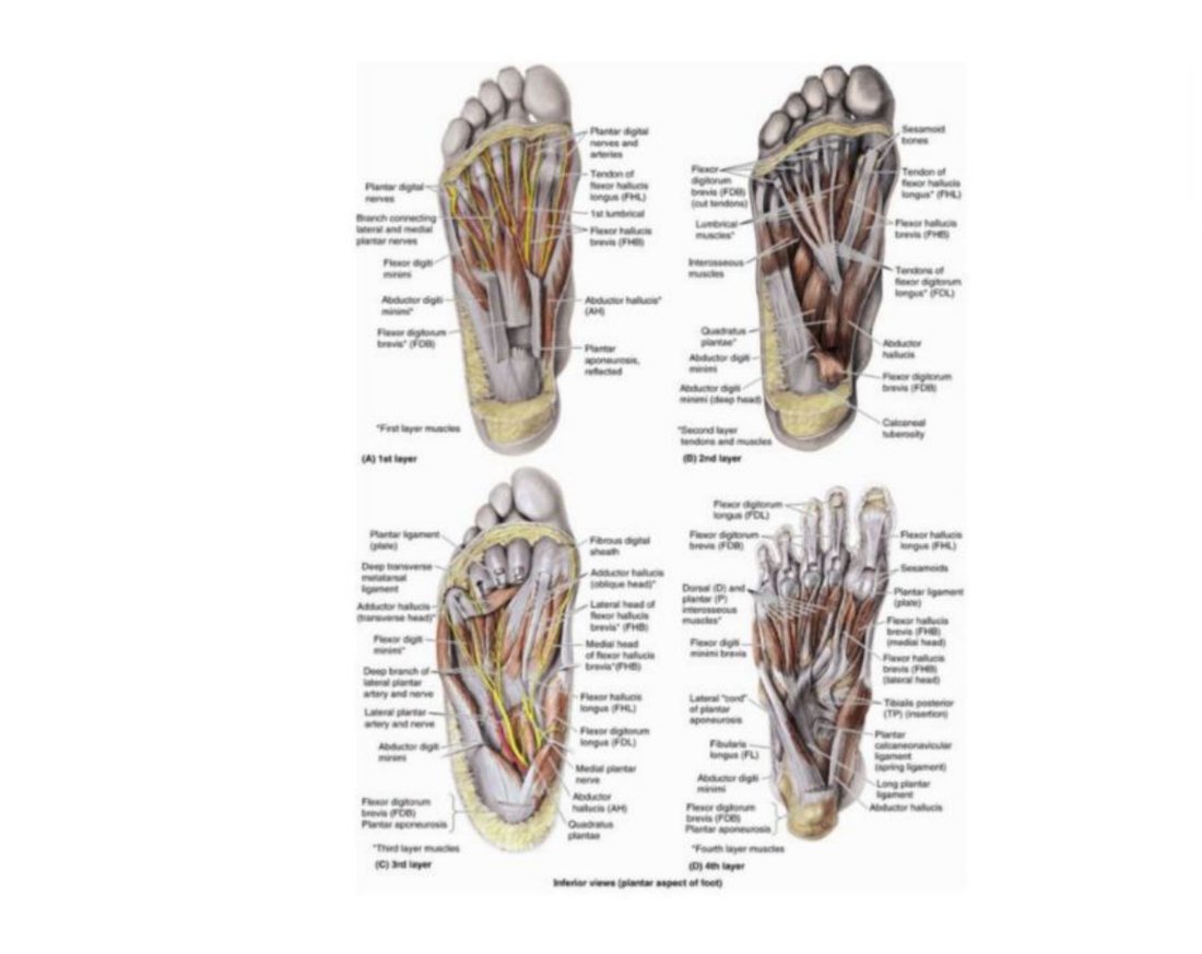
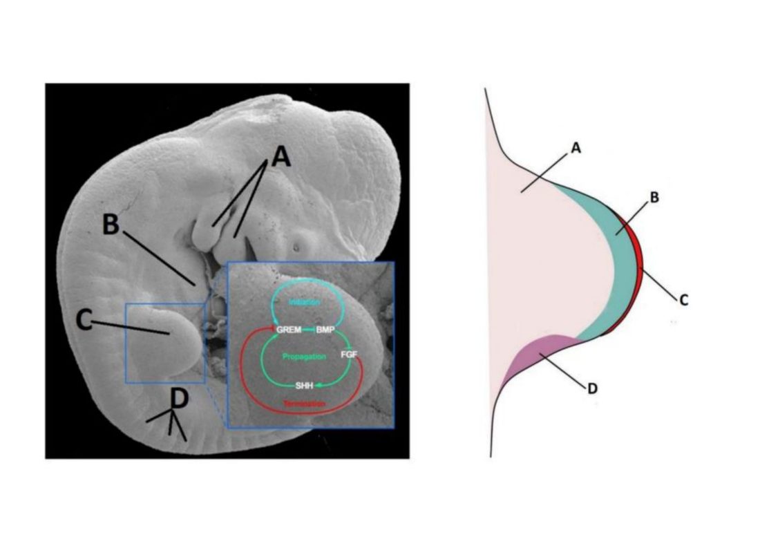
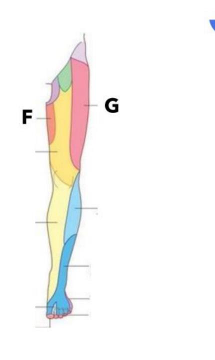
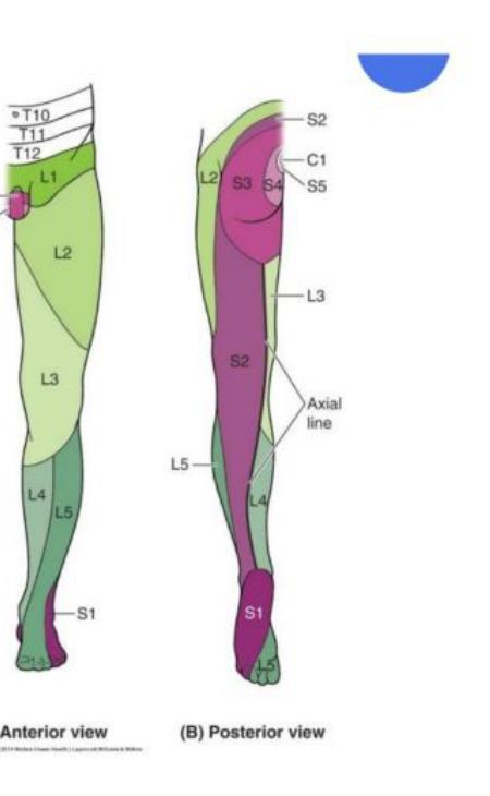
Practice questions on Lower Limb
A patient with a deep venous thrombosis of the lower limb experiences a sudden, massive collapse and fatal cardiovascular failure due to a large thrombus occluding the pulmonary artery bifurcation. What is the classic term used to describe this specific large embolus?
- Marantic vegetation on cardiac valves
- Saddle embolus occluding main bifurcation
- Ghon focus within the lung parenchyma
- Aschoff body found in myocardial tissue
Answer: B. A saddle embolus refers to a large thromboembolus that occludes the bifurcation of the main pulmonary artery, causing sudden death. Marantic vegetations are sterile non-infective endocardial lesions associated with hypercoagulable states.
A 24-year-old medical student is revising lower limb compartments. Which of the following describes the actions of the adductor group of muscles of the thigh?
- Strong adductor and medial rotator of the hip
- Flexor of the hip and abductor of the thigh
- Extensor of the knee and lateral rotator
- Weak adductor and lateral rotator of the hip
Answer: A. The adductor muscle group acts as a strong adductor and a medial rotator of the hip. They do not primarily act as hip flexors and abductors.
During dissection of the lower limb, a student explores the subsartorial (adductor) canal. Which neurovascular structure lies deep to boundary B within this canal?
- Saphenous nerve
- Common peroneal nerve
- Deep fibular nerve
- Lateral cutaneous nerve of thigh
Answer: A. The saphenous nerve, nerve to vastus medialis, and femoral vessels lie deep to the adductor canal roof. The common peroneal nerve is located laterally in the lower limb, far from this canal.
Following a supracondylar femoral fracture, displacement of the distal fragment often occurs due to the direct pull of a specific lower limb muscle. What is a major vascular complication associated with this fracture?
- Injury to the popliteal artery
- Thrombosis of the great saphenous vein
- Laceration of the anterior tibial artery
- Rupture of the femoral circumflex artery
Answer: A. Supracondylar femoral fractures can cause injury to the popliteal artery due to the pull of the gastrocnemius muscle on the fragment. The anterior tibial artery is located too far distally to be directly threatened at the supracondylar level.