Retina
The visual receptors in the central part of the retina (fovea centralis) are mainly rods
What our notes say
- The visual receptors in the central part of the retina (fovea centralis) are mainly rods (cat 2 2014 — Source Paper Awaiting Edit)
- One of the following statements regarding the retina is wrong: (Medical Physiology End of Semester 1 Exam — March 2019 (Past Paper))
- Emmetropia is the normal refractive state of the eye where parallel light rays from distant objects (>6 meters) focus exactly on the retina without accommodation. (Medical Physiology Year Two – Paper Two MCQs)
- Photoreceptors: Detect light and are found in the retina of the eye. (MEDICAL PHYSIOLOGY II ESsays)
- a) The photoreceptors (rods and cones) face backwards, i.e. towards the rear surface of the retina (Medical Physiology End of Semester 1 Exam — March 2019 (Past Paper))
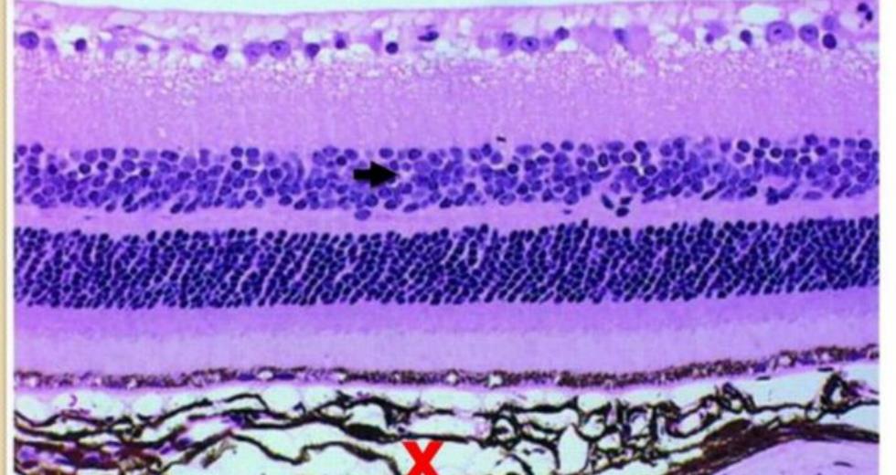
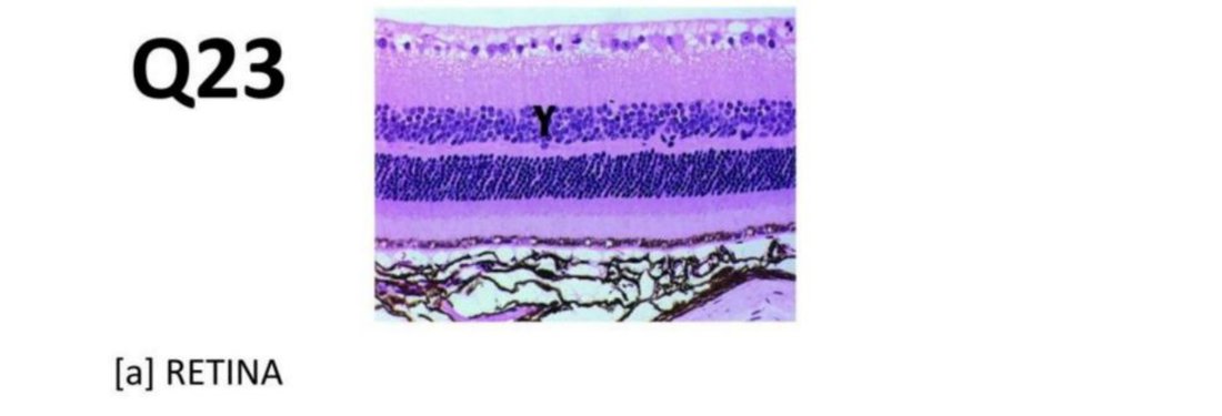
- Retina - recognized by its many neuronal layers and adjacent choroid (Aponeurosis — Histology Question Bank)
Study material on Retina
- Medical Physiology End of Semester 1 Exam — March 2019 (Past Paper)
- Anatomy and Biochemistry of the Eye
- Master First-Year Anatomy: Must-Know Essays & Key Concepts
- cat 2 2014 — Source Paper Awaiting Edit
- Medical Physiology Year Two – Paper Two MCQs
- Sleep Physiology Examination Paper
- Embryology — Past Paper Questions & Answers
Images
Practice questions on Retina
An embryonic eye is developing normally. A retina is present on the posterior aspect of the globe. What is the embryologic origin of the portion of retina containing nerve cell bodies?
- Diencephalon
- Mesoderm
- Notochord
- Pharyngeal arch
- Surface ectoderm
Answer: A. The neural retina is a diencephalic structure. The telencephalon (cerebral hemispheres) and the diencephalon (including thalamus and hypothalamus) are derivatives of the prosencephalon. ---
Cotton wool spots in the retina represent:
- Lipid exudates
- Microaneurysms
- Nerve fibre layer infarcts
- Retinal tears
- Tumour infiltration
Answer: C. Cotton wool spots are soft exudates in the retina that represent microinfarcts of the retinal nerve fiber layer, often seen in conditions like severe hypertension or diabetes.
With regards to the retina, which is CORRECT?
- The retina covers the inner surface of the choroid and is light sensitive everywhere except at the corneal area
- The optic disc contains retina that is completely free of blood vessels and is yellowish in color
- The optic disc and fovea are of similar size
- The fovea contains no blood vessels or cones, but a high concentration of rods
Answer: C. ** The optic disc and fovea are indeed of similar size (approximately 1.5mm in diameter). The fovea contains a high concentration of cones, not rods, and has no blood vessels. ---