Pulmonary Hypertension
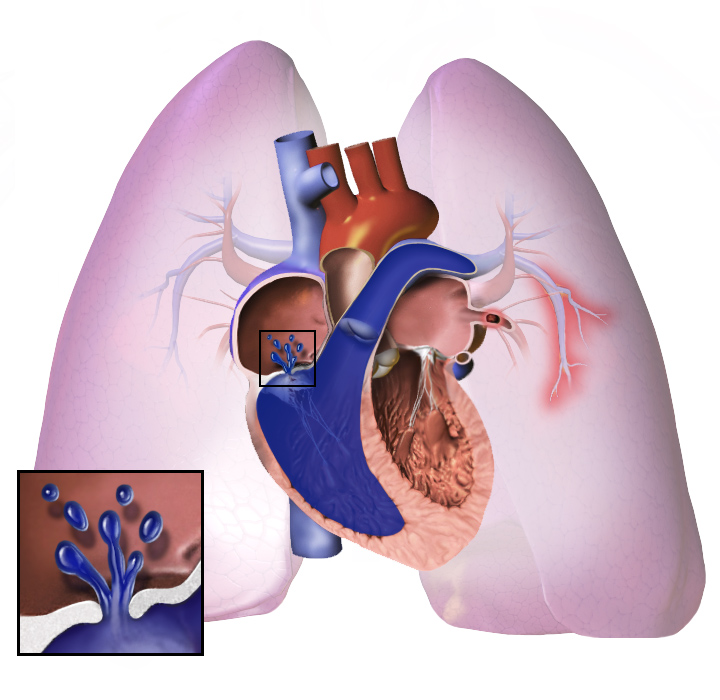
Pulmonary Hypertension (PH) is a haemodynamic and pathophysiological condition defined by an increase in mean pulmonary arterial pressure (mPAP) ≥ 20 mmHg at rest, as measured by right heart catheterisation (RHC).
Pulmonary Hypertension
- Defined as pulmonary pressures of ≥25 mmHg at rest . Normally, pulmonary blood pressures are one-eighth of systemic pressures.
- WHO Classification (5 Groups):
- Pulmonary arterial hypertension : Includes heritable forms, connective tissue diseases, HIV infection, and congenital heart disease with left-to-right shunts.
- Left-sided heart disease : Including systolic/diastolic dysfunction and valvular disease.
- Lung diseases and/or hypoxia : Such as COPD, interstitial lung disease, and obstructive sleep apnea.
Read this in Lung Pathology — Must-Know
What our notes say
- Pulmonary Hypertension (PH) is a haemodynamic and pathophysiological condition defined by an increase in mean pulmonary arterial pressure (mPAP) ≥ 20 mmHg at rest, as measured by right heart catheterisation (RHC). (Pulmonary hypertension and cor pulmonale)
- Result: airflow obstruction, V/Q mismatch (hypoxaemia), hypercapnia in advanced disease, pulmonary hypertension and cor pulmonale. (Chronic Obstructive Pulmonary Disease (COPD))
- Long-term oxygen therapy (LTOT): for PaO2 below 7.3 kPa (55 mmHg), or 7.3–8 kPa with cor pulmonale, polycythaemia or pulmonary hypertension, used at least 15 hours a day; patient must not smoke. (Chronic Obstructive Pulmonary Disease (COPD))
- | Pulmonary vasculature (arterioles & veins) – thin‑walled muscular vessels | Fibrotic distortion narrows vessels, raising pulmonary vascular resistance → secondary pulmonary hypertension and right‑heart strain (cor pulmonale). | (Interstitial Lung Diseases)
- Pulmonary vascular remodeling → pulmonary hypertension → right‑sided heart failure (Interstitial Lung Diseases)
- | Pulmonary hypertension | Chronic hypoxic vasoconstriction + loss of capillary bed from fibrosis | Exertional dyspnoea out of proportion to spirometry, right‑sided edema | Late stage, 10–20 yr after disease onset | (Occupational Lung Disease)
Study material on Pulmonary Hypertension
- Pulmonary hypertension and cor pulmonale
- Lung Pathology — Must-Know
- Respiratory System Pathology 1A
- Cardiovascular system examination revised 2012
- Interstitial Lung Diseases
- CARDIOVASCULAR PATHOLOGY
- CARDIOVASCULAR PATHOLOGY
- Heart Disease: Must Knows
- Heart failure: which tests and why
- Sickle Cell Disease: Pathology, Genetics & Mechanisms
- Respiratory Pathology
Images
Practice questions on Pulmonary Hypertension
A 58-year-old man develops DVT postoperatively then has a left MCA stroke. CXR shows pulmonary hypertension. Troponin normal. Most likely lesion on echo?
- Coarctation of the aorta
- Tetralogy of Fallot
- Atrial septal defect
- Pulmonic stenosis
Answer: C. DVT → paradoxical embolism through an ASD → systemic arterial stroke. Pulmonary hypertension supports a long-standing shunt that has reversed (Eisenmenger complex).
In pulmonary hypertension, plexiform lesions are characteristic of which type?
- PH secondary to mitral stenosis
- PH secondary to recurrent pulmonary embolism
- Idiopathic (primary) pulmonary arterial hypertension
- PH secondary to chronic lung disease
Answer: C. Plexiform lesions — endothelial proliferation forming multiple lumina within small arteries — are characteristic of idiopathic (primary) pulmonary arterial hypertension, not secondary forms.
The BMPR-2 gene mutation in primary pulmonary hypertension leads to
- Increased vasodilator production by endothelial cells
- Abnormal endothelial and smooth muscle cell proliferation
- Increased serotonin transporter expression on smooth muscle
- Enhanced prostacyclin synthesis in pulmonary arteries
Answer: B. BMPR-2 normally inhibits cell proliferation. Loss-of-function mutations found in 50% of familial primary pulmonary hypertension cases result in abnormal vascular endothelial and smooth muscle proliferation.
A young man uses intravenous drugs and develops pulmonary hypertension. Lung biopsy shows granulomas with birefringent crystals under polarized light. The likely substance is
- Silica particles from contaminated needles
- Asbestos fibers from environmental exposure
- Magnesium trisilicate (talc) from IV drug mixture
- Coal dust from occupational inhalation
Answer: C. IV drug abuse introduces magnesium trisilicate (talc) as a filler in drug mixtures into the pulmonary microvasculature. Talc elicits a granulomatous response; vascular involvement leads to pulmonary hypertension. Talc crystals are visible by polarized light.