Heart
Consider if the patient is systemically unwell, very frail, has comorbidity (COPD, heart failure), or pertussis is suspected (macrolide).
Treatment at a glance
- ACE inhibitor or ARB (or ARNI)
- Beta-blocker (bisoprolol, carvedilol)
- Mineralocorticoid antagonist (spironolactone)
What our notes say
- Consider if the patient is systemically unwell, very frail, has comorbidity (COPD, heart failure), or pertussis is suspected (macrolide). (Bronchitis (Acute and Chronic))
- Side effects: reduced appetite, insomnia, increased heart rate and blood pressure, slowed growth in children (monitor height and weight), potential for misuse. (Introduction to Psychopharmacology)
- | Medically ill patients | Adjust for kidney and liver disease; check heart disease and QT prolongation; consider interactions with other drugs. | (Introduction to Psychopharmacology)
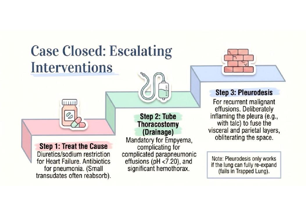
- Review at 48 hours; if no improvement, think of empyema, resistant or unusual organism, TB, obstruction (cancer), wrong diagnosis (PE, heart failure). (Pneumonia)
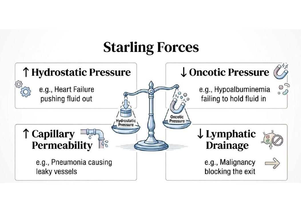
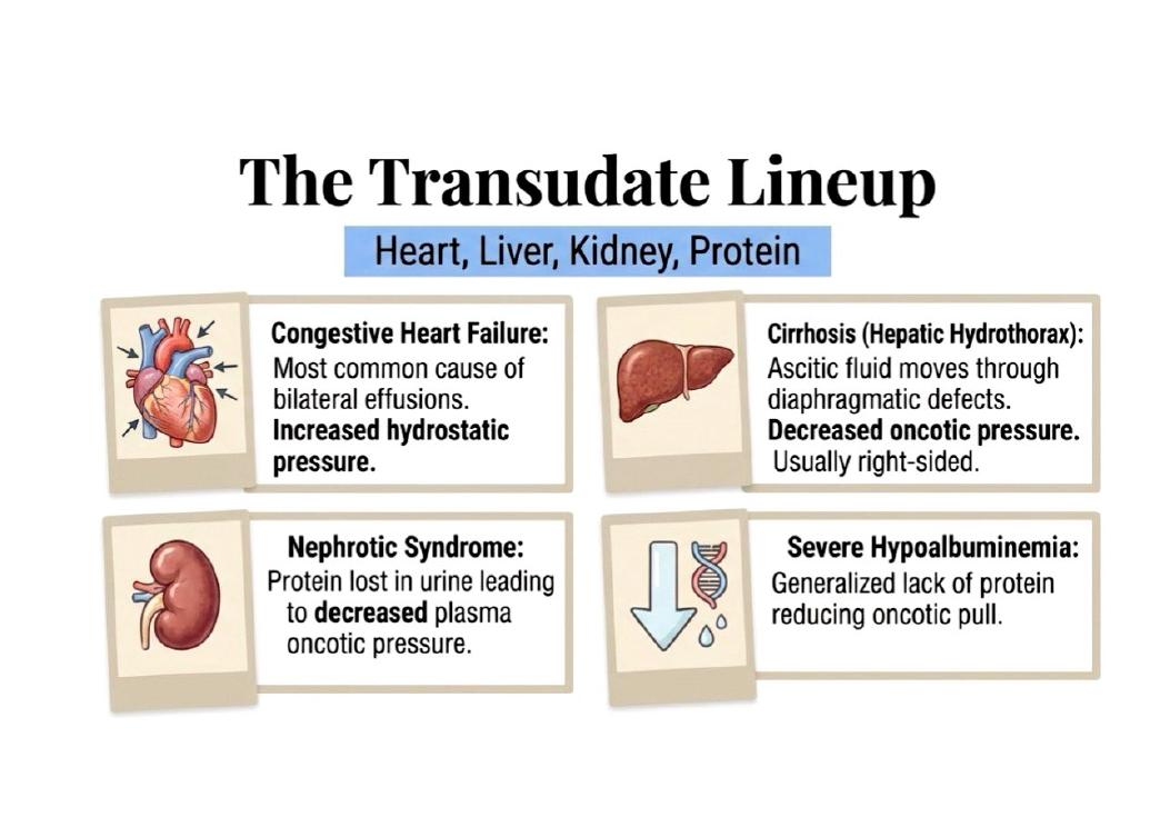
- Increased hydrostatic pressure (heart failure) → transudate. (Pleural Effusion)
- | Heart failure (commonest, bilateral) | Pneumonia (parapneumonic effusion, empyema) | (Pleural Effusion)
Study material on Heart
- Cardiovascular system examination revised 2012
- Heart Disease — Must Knows
- Heart Disease: Must Knows
- 🫀 VALVULAR HEART DISEASE — ULTRA DEEP NOTES
- Comprehensive Heart Pathology Notes 🫀
- Heart Pathology: Comprehensive Short Notes
- Heart failure: which tests and why
- Acute rheumatic fever and rheumatic heart disease
- Paediatrics end of 3rd trimester exam (MBPE 4300): 11 January 2022, congenital heart disease essay and 20 MCQs
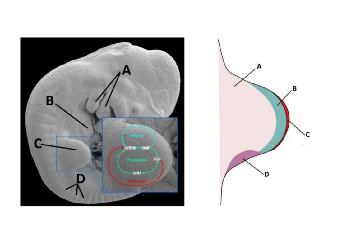
- Development of the Heart
- Heart Disease — Must Knows
- Heart Disease: Must Knows
- Heart Disease: Must Knows
- Congenital Heart Disease
- Congenital Heart Disease
- Congenital Heart Disease
Images

Practice questions on Heart
A 55-year-old man with heart failure has a right-sided pleural effusion. Thoracentesis shows fluid protein of 2.1 g/dL, serum protein 7.0 g/dL, fluid LDH 88 U/L, and serum LDH 300 U/L. What is the most likely type of effusion?
- Exudative
- Transudative
- Chylous
- Hemorrhagic
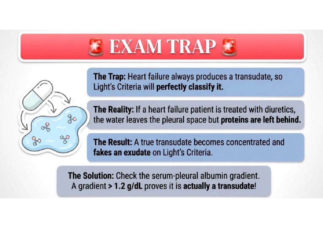
Answer: B. Using Light's criteria, this is a transudative effusion: Fluid protein/serum protein ratio is 2.1/7.0 = 0.3 ( < 0.5), and fluid LDH/serum LDH ratio is 88/300 = 0.29 (< 0.6). Heart failure is a common cause of transudative effusions.
A 32-year-old woman with no cardiopulmonary disease has progressive exertional dyspnea, syncope on exertion, and loud P2 on auscultation. Right heart catheterization shows mean pulmonary artery pressure of 38 mmHg with normal pulmonary capillary wedge pressure. What is the most likely diagnosis?
- Mitral stenosis
- Pulmonary arterial hypertension (PAH)
- Left ventricular failure
- Pulmonary embolism
Answer: B. The combination of exertional dyspnea, syncope, loud P2, elevated pulmonary artery pressure, and normal pulmonary capillary wedge pressure strongly suggests pulmonary arterial hypertension (PAH).
A 16-year-old is stabbed in the left chest. BP barely obtainable, lungs clear, heart sounds barely audible. What is the most useful treatment?
- Intravenous fluids and observation
- Needle decompression
- Pericardiocentesis
- Tube thoracostomy
Answer: D. The patient's hemodynamic instability (barely obtainable BP) and clear lungs with faint heart sounds after chest trauma strongly suggest cardiac tamponade. Tube thoracostomy is indicated to drain the pericardial space.
An infant born at term has a radiograph taken. No congenital abnormalities are noted. The upper heart borders are not well visualized. Which of the following structures normally present is most likely to obscure the heart borders in this infant's radiograph?
- Esophagus
- Lymph node
- Stomach
- Thymus
- Vertebra
Answer: D. The thymus is in the anterior mediastinum and is proportionately larger in a newborn than later in life, and is busily schooling T lymphocytes, so it has a soft tissue density similar to the heart that lies posterior to it. In adults, the thymus appears on radiographs as only a small fatty remnant. ---
A 1-month-old infant has failure to thrive. On examination a heart murmur is auscultated over the chest. No cyanosis is noted. Echocardiography shows a left-to-right shunt just above the muscular interventricular septum. An abnormality of which of the following structures most likely produced this infant's defect?
- Chordae tendineae
- Crista terminalis
- Endocardial cushion
- Septum primum
- Sinus venosus
Answer: C. The endocardial cushions contribute to the atrioventricular valves and the septae,including the membranous portion of the interventricular septum. This is the most common form of ventricular septal defect (VSD), which is the most common heart defect in liveborns. ---
50-year-old, sudden cardiac death, heart 560g, concentric LV wall thickening, normal-sized chamber. Most likely hemodynamic cause?
- Chronic aortic regurgitation
- Chronic systemic hypertension (pressure overload)
- Large ventricular septal defect (volume overload)
- Chronic mitral regurgitation
- Tricuspid stenosis
Answer: B. - **Concentric hypertrophy with normal chamber size = pressure overload** (hypertension, aortic stenosis) - Volume overload lesions (MR, AR, VSD) cause **eccentric** hypertrophy with chamber **dilation** ---